
Rabbit Polyclonal Antibody to Phospho-MYT1(T495)
货号:
P30151
别名:
Membrane-associated tyrosine- and threonine-specific cdc2-inhibitory kinase, Myt1 kinase, PKMYT1, MYT1
应用:
WB,IHC
反应种属:
Human
抗体类型:
Primary antibody
Swissprot:
Q99640
规格:
目录价
在线咨询
Description |
|---|
The protein encoded by this gene is a member of the serine/threonine protein kinase family. This kinase preferentially phosphorylates and inactivates cell division cycle 2 protein (CDC2), and thus negatively regulates cell cycle G2/M transition. This kinase is associated with the membrane throughout the cell cycle. Its activity is highly regulated during the cell cycle. Protein kinases AKT1/PKB and PLK (Polo-like kinase) have been shown to phosphorylate and regulate the activity of this kinase. Alternatively spliced transcript variants encoding distinct isoforms have been reported. |
Specification |
|
|---|---|
| Aliases | Membrane-associated tyrosine- and threonine-specific cdc2-inhibitory kinase, Myt1 kinase, PKMYT1, MYT1 |
| Entrez GeneID | 9088 |
| Swissprot | Q99640 |
| WB Predicted band size | 54.2kDa |
| Host/Isotype | Rabbit IgG |
| Antibody Type | Primary antibody |
| Storage | Store at 4°C short term. Aliquot and store at -20°C long term. Avoid freeze/thaw cycles. |
| Species Reactivity | Human |
| Immunogen | This MYT1 Antibody is generated from rabbits immunized with a KLH conjugated synthetic phosphopeptide corresponding to amino acid residues surrounding T495 of human MYT1. |
| Formulation | Purified antibody in PBS with 0.05% sodium azide. |
Application |
|
|---|---|
| WB | 1/500-1/1000 |
| IHC | 1/100-1/500 |
Product Image
-
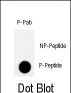
Dot blot analysis of anti-Phospho-MYT1-T495 Antibody (Cat. #P30151) on nitrocellulose membrane. 50ng of Phospho-peptide or Non Phospho-peptide per dot were adsorbed. Antibody working concentrations are 0.5ug per ml.

-
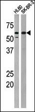
The anti-Phospho-MYT1-T495 Antibody (Cat. #P30151) is used in Western blot to detect Phospho-MYT1-T495 in HL60 (left) and SK-BR-3 (right) tissue lysates.

-
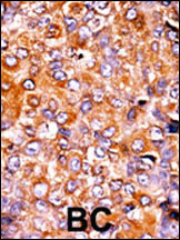
Formalin-fixed and paraffin-embedded human cancer tissue reacted with the primary antibody, which was peroxidase-conjugated to the secondary antibody, followed by AEC staining. This data demonstrates the use of this antibody for immunohistochemistry; clinical relevance has not been evaluated. BC = breast carcinoma; HC = hepatocarcinoma.


 鄂公网安备42018502007531号

