
Mouse Monoclonal Antibody to PINK1
货号:
P30246
别名:
Serine/threonine-protein kinase PINK1, mitochondrial, BRPK, PTEN-induced putative kinase protein 1, PINK1
应用:
WB,IHC
反应种属:
Human, Mouse
抗体类型:
Primary antibody
Swissprot:
Q9BXM7
规格:
目录价
在线咨询
Description |
|---|
This gene encodes a serine/threonine protein kinase that localizes to mitochondria. It is thought to protect cells from stress-induced mitochondrial dysfunction. Mutations in this gene cause one form of autosomal recessive early-onset Parkinson disease. |
Specification |
|
|---|---|
| Aliases | Serine/threonine-protein kinase PINK1, mitochondrial, BRPK, PTEN-induced putative kinase protein 1, PINK1 |
| Entrez GeneID | 65018 |
| Swissprot | Q9BXM7 |
| WB Predicted band size | 62.8kDa |
| Host/Isotype | Mouse IgG1 |
| Antibody Type | Primary antibody |
| Storage | Store at 4°C short term. Aliquot and store at -20°C long term. Avoid freeze/thaw cycles. |
| Species Reactivity | Human, Mouse |
| Immunogen | Recombinant PINK1 protein was used to produced this monoclonal antibody. |
| Formulation | Purified antibody in PBS with 0.05% sodium azide. |
Application |
|
|---|---|
| WB | 1/500-1/2000 |
| IHC | 1/100-1/500 |
Product Image
-
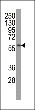
Western blot analysis of anti-PINK1 Monoclonal Antibody (P30246) in mouse brain tissue lysates. PINK1(arrow) was detected using the ascites Mab. (dilution 1:500)

-
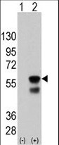
Western blot analysis of PINK (arrow) using mouse monoclonal PINK antibody(Ascites). 293 cell lysates (2 μg/lane) either nontransfected (Lane 1) or transiently transfected with the PINK gene (Lane 2) (Origene Technologies) (1:2000)

-
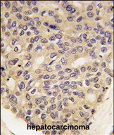
Formalin-fixed and paraffin-embedded human hepatocarcinoma tissue reacted with PINK1 Monoclonal Antibody (Cat.#P30246), which was peroxidase-conjugated to the secondary antibody, followed by DAB staining. This data demonstrates the use of this antibody for immunohistochemistry; clinical relevance has not been evaluated.

-
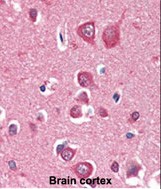
Formalin-fixed and paraffin-embedded human Brain Cortex tissue reacted with PINK1 Monoclonal Antibody (Cat.#P30246), which was peroxidase-conjugated to the secondary antibody, followed by AEC staining. This data demonstrates the use of this antibody for immunohistochemistry; clinical relevance has not been evaluated.


 鄂公网安备42018502007531号

