
Rabbit Polyclonal Antibody to GCAT
货号:
P30356
别名:
2-amino-3-ketobutyrate coenzyme A ligase, mitochondrial, AKB ligase, Aminoacetone synthase, Glycine acetyltransferase, GCAT, KBL
应用:
WB,IHC,FCM
反应种属:
Human, Mouse
抗体类型:
Primary antibody
Swissprot:
O75600
规格:
目录价
在线咨询
Description |
|---|
The degradation of L-threonine to glycine consists of a two-step biochemical pathway involving the enzymes L-threonine dehydrogenase and 2-amino-3-ketobutyrate coenzyme A ligase. L-Threonine is first converted into 2-amino-3-ketobutyrate by L-threonine dehydrogenase. GCAT is the second enzyme in this pathway, which then catalyzes the reaction between 2-amino-3-ketobutyrate and coenzyme A to form glycine and acetyl-CoA. The enzyme is considered a class II pyridoxal-phosphate-dependent aminotransferase. |
Specification |
|
|---|---|
| Aliases | 2-amino-3-ketobutyrate coenzyme A ligase, mitochondrial, AKB ligase, Aminoacetone synthase, Glycine acetyltransferase, GCAT, KBL |
| Entrez GeneID | 23464 |
| Swissprot | O75600 |
| WB Predicted band size | 45.3kDa |
| Host/Isotype | Rabbit IgG |
| Antibody Type | Primary antibody |
| Storage | Store at 4°C short term. Aliquot and store at -20°C long term. Avoid freeze/thaw cycles. |
| Species Reactivity | Human, Mouse |
| Immunogen | This GCAT antibody is generated from rabbits immunized with a KLH conjugated synthetic peptide between 155-181 amino acids from the Central region of human GCAT. |
| Formulation | Purified antibody in PBS with 0.05% sodium azide,1%BSA and 50% glycerol.prepared by Saturated Ammonium Sulfate (SAS) . |
Application |
|
|---|---|
| WB | 1/1000 |
| IHC | 1/100-1/500 |
| FCM | 1/10-1/50 |
Product Image
-
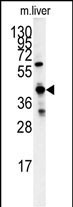
Western blot analysis of GCAT antibody (Center) (Cat.#P30356) in mouse liver tissue lysates (35ug/lane). GCAT (arrow) was detected using the purified Pab.

-
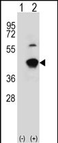
Western blot analysis of GCAT (arrow) using rabbit polyclonal GCAT Antibody (Center) (Cat.#P30356). 293 cell lysates (2 ug/lane) either nontransfected (Lane 1) or transiently transfected (Lane 2) with the GCAT gene.

-
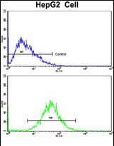
Flow cytometric analysis of HepG2 cells using GCAT Antibody (Center)(bottom histogram) compared to a negative control cell (top histogram). FITC-conjugated goat-anti-rabbit secondary antibodies were used for the analysis.

-
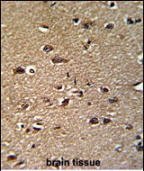
Formalin-fixed and paraffin-embedded human brain reacted with GCAT Antibody (Center), which was peroxidase-conjugated to the secondary antibody, followed by DAB staining. This data demonstrates the use of this antibody for immunohistochemistry; clinical relevance has not been evaluated.


 鄂公网安备42018502007531号

