
Rabbit Polyclonal Antibody to VIP
货号:
P30377
别名:
VIP peptides, Intestinal peptide PHV-42, Peptide histidine valine 42, Intestinal peptide PHM-27, Peptide histidine methioninamide 27, Vasoactive intestinal peptide, VIP, Vasoactive intestinal polypeptide, VIP
应用:
WB,IHC,FCM
反应种属:
Human
抗体类型:
Primary antibody
Swissprot:
P01282
规格:
目录价
在线咨询
Description |
|---|
VIP belongs to the glucagon family. It stimulates myocardial contractility, causes vasodilation, increases glycogenolysis, lowers arterial blood pressure and relaxes the smooth muscle of trachea, stomach and gall bladder. |
Specification |
|
|---|---|
| Aliases | VIP peptides, Intestinal peptide PHV-42, Peptide histidine valine 42, Intestinal peptide PHM-27, Peptide histidine methioninamide 27, Vasoactive intestinal peptide, VIP, Vasoactive intestinal polypeptide, VIP |
| Entrez GeneID | 7432 |
| Swissprot | P01282 |
| WB Predicted band size | 19.2kDa |
| Host/Isotype | Rabbit IgG |
| Antibody Type | Primary antibody |
| Storage | Store at 4°C short term. Aliquot and store at -20°C long term. Avoid freeze/thaw cycles. |
| Species Reactivity | Human |
| Immunogen | This VIP antibody is generated from rabbits immunized with a KLH conjugated synthetic peptide between 139-167 amino acids from the C-terminal region of human VIP. |
| Formulation | Purified antibody in PBS with 0.05% sodium azide,1%BSA and 50% glycerol.prepared by Saturated Ammonium Sulfate (SAS) . |
Application |
|
|---|---|
| WB | 1/1000 |
| IHC | 1/100-1/500 |
| FCM | 1/10-1/50 |
Product Image
-
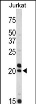
Western blot analysis of VIP antibody (C-term) (Cat. #P30377) in Jurkat cell line lysates (35ug/lane). VIP (arrow) was detected using the purified Pab.

-
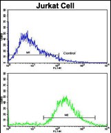
Flow cytometric analysis of jurkat cells using VIP Antibody (C-term)(bottom histogram) compared to a negative control cell (top histogram). FITC-conjugated goat-anti-rabbit secondary antibodies were used for the analysis.

-
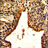
Formalin-fixed and paraffin-embedded human prostate carcinoma with VIP Antibody (C-term), which was peroxidase-conjugated to the secondary antibody, followed by DAB staining. This data demonstrates the use of this antibody for immunohistochemistry; clinical relevance has not been evaluated.


 鄂公网安备42018502007531号

