
Rabbit Polyclonal Antibody to AMT (N-term)
货号:
P30401
别名:
Aminomethyltransferase, mitochondrial, Glycine cleavage system T protein, GCVT, AMT, GCST
应用:
WB,IHC,FCM
反应种属:
Human
抗体类型:
Primary antibody
Swissprot:
P48728
规格:
目录价
在线咨询
Description |
|---|
The enzyme system for cleavage of glycine (glycine cleavage system; EC 2.1.2.10), which is confined to the mitochondria, is composed of 4 protein components: P protein (a pyridoxal phosphate-dependent glycine decarboxylase; MIM 238300), H protein (a lipoic acid-containing protein; MIM 238330), T protein (a tetrahydrofolate-requiring enzyme), and L protein (a lipoamide dehydrogenase; MIM 238331). Glycine encephalopathy (GCE; MIM 605899) may be due to a defect in any one of these enzymes. |
Specification |
|
|---|---|
| Aliases | Aminomethyltransferase, mitochondrial, Glycine cleavage system T protein, GCVT, AMT, GCST |
| Entrez GeneID | 275 |
| Swissprot | P48728 |
| WB Predicted band size | 43.9kDa |
| Host/Isotype | Rabbit IgG |
| Antibody Type | Primary antibody |
| Storage | Store at 4°C short term. Aliquot and store at -20°C long term. Avoid freeze/thaw cycles. |
| Species Reactivity | Human |
| Immunogen | This AMT antibody is generated from rabbits immunized with a KLH conjugated synthetic peptide between 19-45 amino acids from the N-terminal region of human AMT. |
| Formulation | Purified antibody in PBS with 0.05% sodium azide,1%BSA and 50% glycerol.prepared by Saturated Ammonium Sulfate (SAS) . |
Application |
|
|---|---|
| WB | 1/1000 |
| IHC | 1/100-1/500 |
| FCM | 1/10-1/50 |
Product Image
-
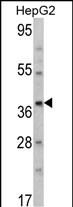
Western blot analysis of AMT Antibody (N-term) (Cat. #P30401) in HepG2 cell line lysates (35ug/lane). AMT (arrow) was detected using the purified Pab.

-
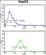
AMT Antibody (N-term) (Cat.#P30401) flow cytometry analysis of HepG2 cells (bottom histogram) compared to a negative control cell (top histogram). FITC-conjugated goat-anti-rabbit secondary antibodies were used for the analysis.

-
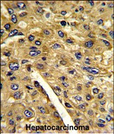
Formalin-fixed and paraffin-embedded human hepatocarcinoma reacted with AMT Antibody (N-term), which was peroxidase-conjugated to the secondary antibody, followed by DAB staining. This data demonstrates the use of this antibody for immunohistochemistry; clinical relevance has not been evaluated.


 鄂公网安备42018502007531号

