
Rabbit Polyclonal Antibody to TSPAN6
货号:
P30476
别名:
Tetraspanin-6, Tspan-6, A15 homolog, Putative NF-kappa-B-activating protein 321, T245 protein, Tetraspanin TM4-D, Transmembrane 4 superfamily member 6, TSPAN6, TM4SF6
应用:
WB,FCM
反应种属:
Human, Mouse
抗体类型:
Primary antibody
Swissprot:
O43657
规格:
目录价
在线咨询
Description |
|---|
The protein is a member of the transmembrane 4 superfamily, also known as the tetraspanin family. Most of these members are cell-surface proteins that are characterized by the presence of four hydrophobic domains. The proteins mediate signal transduction events that play a role in the regulation of cell development, activation, growth and motility. This encoded protein is a cell surface glycoprotein and is highly similar in sequence to the transmembrane 4 superfamily member 2. |
Specification |
|
|---|---|
| Aliases | Tetraspanin-6, Tspan-6, A15 homolog, Putative NF-kappa-B-activating protein 321, T245 protein, Tetraspanin TM4-D, Transmembrane 4 superfamily member 6, TSPAN6, TM4SF6 |
| Entrez GeneID | 7105 |
| Swissprot | O43657 |
| WB Predicted band size | 27.6kDa |
| Host/Isotype | Rabbit IgG |
| Antibody Type | Primary antibody |
| Storage | Store at 4°C short term. Aliquot and store at -20°C long term. Avoid freeze/thaw cycles. |
| Species Reactivity | Human, Mouse |
| Immunogen | This TSPAN6 antibody is generated from rabbits immunized with a KLH conjugated synthetic peptide between 176~205 amino acids from the C-terminal region of human TSPAN6. |
| Formulation | Purified antibody in PBS with 0.05% sodium azide. |
Application |
|
|---|---|
| WB | 1/1000 |
| FCM | 1/10-1/50 |
Product Image
-
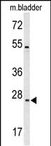
Western blot analysis of TSPAN6 Antibody (C-term) (Cat. #P30476) in mouse bladder tissue lysates (35ug/lane). TSPAN6 (arrow) was detected using the purified Pab.

-
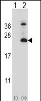
Western blot analysis of TSPAN6 (arrow) using rabbit polyclonal TSPAN6 Antibody (C-term) (Cat. #P30476). 293 cell lysates (2 ug/lane) either nontransfected (Lane 1) or transiently transfected (Lane 2) with the TSPAN6 gene.

-
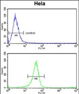
TSPAN6 Antibody (C-term) (Cat. #P30476) flow cytometry analysis of Hela cells (bottom histogram) compared to a negative control cell (top histogram).FITC-conjugated goat-anti-rabbit secondary antibodies were used for the analysis.


 鄂公网安备42018502007531号

