
Rabbit Polyclonal Antibody to PGD
货号:
P30681
别名:
6-phosphogluconate dehydrogenase, decarboxylating, PGD, PGDH
应用:
WB,IHC,FCM
反应种属:
Human, Mouse
抗体类型:
Primary antibody
Swissprot:
P52209
规格:
目录价
在线咨询
Description |
|---|
6-phosphogluconate dehydrogenase is the second dehydrogenase in the pentose phosphate shunt. Deficiency of this enzyme is generally asymptomatic, and the inheritance of this disorder is autosomal dominant. Hemolysis results from combined deficiency of 6-phosphogluconate dehydrogenase and 6-phosphogluconolactonase suggesting a synergism of the two enzymopathies. |
Specification |
|
|---|---|
| Aliases | 6-phosphogluconate dehydrogenase, decarboxylating, PGD, PGDH |
| Entrez GeneID | 5226 |
| Swissprot | P52209 |
| WB Predicted band size | 53.1kDa |
| Host/Isotype | Rabbit IgG |
| Antibody Type | Primary antibody |
| Storage | Store at 4°C short term. Aliquot and store at -20°C long term. Avoid freeze/thaw cycles. |
| Species Reactivity | Human, Mouse |
| Immunogen | This PGD antibody is generated from rabbits immunized with a KLH conjugated synthetic peptide between 236-265 amino acids from the Central region of human PGD. |
| Formulation | Purified antibody in PBS with 0.05% sodium azide. |
Application |
|
|---|---|
| WB | 1/1000 |
| IHC | 1/100-1/500 |
| FCM | 1/10-1/50 |
Product Image
-
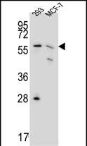
PGD Antibody (Center) (Cat.#P30681) western blot analysis in 293,MCF-7 cell line lysates (35ug/lane).This demonstrates the PGD antibody detected PGD protein (arrow).

-
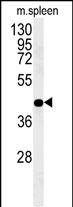
PGD Antibody (Center) (Cat.#P30681) western blot analysis in mouse spleen tissue lysates (35ug/lane).This demonstrates the PGD antibody detected PGD protein (arrow).

-
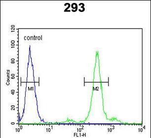
PGD Antibody (Center) (Cat. #P30681) flow cytometric analysis of 293 cells (right histogram) compared to a negative control cell (left histogram).FITC-conjugated goat-anti-rabbit secondary antibodies were used for the analysis.

-
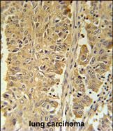
PGD Antibody (Center) (Cat. #P30681) immunohistochemistry analysis in formalin fixed and paraffin embedded human lung carcinoma followed by peroxidase conjugation of the secondary antibody and DAB staining. This data demonstrates the use of the PGD Antibody (Center) for immunohistochemistry. Clinical relevance has not been evaluated.


 鄂公网安备42018502007531号

