
Rabbit Polyclonal Antibody to GOLGA5
货号:
P31405
别名:
Golgin subfamily A member 5, Cell proliferation-inducing gene 31 protein, Golgin-84, Protein Ret-II, RET-fused gene 5 protein, GOLGA5, RETII, RFG5
应用:
WB,FCM
反应种属:
Human
抗体类型:
Primary antibody
Swissprot:
Q8TBA6
规格:
目录价
在线咨询
Description |
|---|
The Golgi apparatus, which participates in glycosylation and transport of proteins and lipids in the secretory pathway, consists of a series of stacked cisternae (flattened membrane sacs). Interactions between the Golgi and microtubules are thought to be important for the reorganization of the Golgi after it fragments during mitosis. This gene encodes one of the golgins, a family of proteins localized to the Golgi. This protein is a coiled-coil membrane protein that has been postulated to play a role in vesicle tethering and docking. Translocations involving this gene and the ret proto-oncogene have been found in tumor tissues; the chimeric sequences have been designated RET-II and PTC5. |
Specification |
|
|---|---|
| Aliases | Golgin subfamily A member 5, Cell proliferation-inducing gene 31 protein, Golgin-84, Protein Ret-II, RET-fused gene 5 protein, GOLGA5, RETII, RFG5 |
| Entrez GeneID | 9950 |
| Swissprot | Q8TBA6 |
| WB Predicted band size | 83.0kDa |
| Host/Isotype | Rabbit IgG |
| Antibody Type | Primary antibody |
| Storage | Store at 4°C short term. Aliquot and store at -20°C long term. Avoid freeze/thaw cycles. |
| Species Reactivity | Human |
| Immunogen | This GOLGA5 antibody is generated from rabbits immunized with a KLH conjugated synthetic peptide between 381-408 amino acids from the Central region of human GOLGA5. |
| Formulation | Purified antibody in PBS with 0.05% sodium azide. |
Application |
|
|---|---|
| WB | 1/1000 |
| FCM | 1/10-1/50 |
Product Image
-
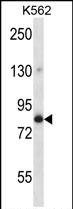
GOLGA5 Antibody (Center) (Cat. #P31405) western blot analysis in K562 cell line lysates (35ug/lane).This demonstrates the GOLGA5 antibody detected the GOLGA5 protein (arrow).

-
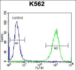
GOLGA5 Antibody (Center) (Cat. #P31405) flow cytometric analysis of K562 cells (right histogram) compared to a negative control cell (left histogram).FITC-conjugated donkey-anti-rabbit secondary antibodies were used for the analysis.


 鄂公网安备42018502007531号

