
Rabbit Polyclonal Antibody to FANCC
货号:
P32583
别名:
Fanconi anemia group C protein, Protein FACC, FANCC, FAC, FACC
应用:
WB,IF
反应种属:
Human
抗体类型:
Primary antibody
Swissprot:
Q00597
规格:
目录价
在线咨询
Description |
|---|
The Fanconi anemia complementation group (FANC) currently includes FANCA, FANCB, FANCC, FANCD1 (also called BRCA2), FANCD2, FANCE, FANCF, FANCG, FANCI, FANCJ (also called BRIP1), FANCL, FANCM and FANCN (also called PALB2). The previously defined group FANCH is the same as FANCA. Fanconi anemia is a genetically heterogeneous recessive disorder characterized by cytogenetic instability, hypersensitivity to DNA crosslinking agents, increased chromosomal breakage, and defective DNA repair. The members of the Fanconi anemia complementation group do not share sequence similarity; they are related by their assembly into a common nuclear protein complex. This protein is for complementation group C. |
Specification |
|
|---|---|
| Aliases | Fanconi anemia group C protein, Protein FACC, FANCC, FAC, FACC |
| Entrez GeneID | 2176 |
| Swissprot | Q00597 |
| WB Predicted band size | 63.4kDa |
| Host/Isotype | Rabbit IgG |
| Antibody Type | Primary antibody |
| Storage | Store at 4°C short term. Aliquot and store at -20°C long term. Avoid freeze/thaw cycles. |
| Species Reactivity | Human |
| Immunogen | This FANCC antibody is generated from rabbits immunized with a KLH conjugated synthetic peptide between 527-555 amino acids from the C-terminal region of human FANCC. |
| Formulation | Purified antibody in PBS with 0.05% sodium azide. |
Application |
|
|---|---|
| WB | 1/1000 |
| IF | 1/10-1/50 |
Product Image
-
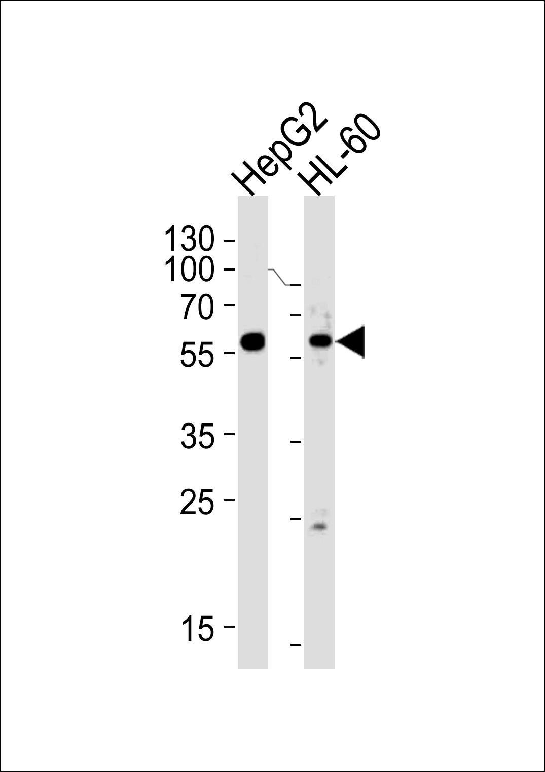
Western blot analysis of lysates from HepG2,HL-60 cell line (from left to right),using FANCC Antibody (C-term)(Cat. #P32583).P32583 was diluted at 1:1000 at each lane. A goat anti-rabbit IgG H&L(HRP) at 1:5000 dilution was used as the secondary antibody.Lysates at 35ug per lane.

-
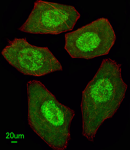
Immunofluorescent analysis of U251 cells, using FANCC Antibody (C-term) (Cat. #P32583). P32583 was diluted at 1:25 dilution. Alexa Fluor 488-conjugated goat anti-rabbit lgG at 1:400 dilution was used as the secondary antibody (green). DAPI was used to stain the cell nuclear (blue).


 鄂公网安备42018502007531号

