
Rabbit Polyclonal Antibody to SAR1A
货号:
P32565
别名:
GTP-binding protein SAR1a, COPII-associated small GTPase, SAR1A, SAR1, SARA, SARA1
应用:
WB,IHC,FCM
反应种属:
Human, Mouse
抗体类型:
Primary antibody
Swissprot:
Q9NR31
规格:
目录价
在线咨询
Description |
|---|
SAR1A is involved in transport from the endoplasmic reticulum to the Golgi apparatus (By similarity) and required to maintain SEC16A localization at discrete locations on the ER membrane perhaps by preventing its dissociation. SAR1A-GTP-dependent assembly of SEC16A on the ER membrane forms an organized scaffold defining endoplasmic reticulum exit sites (ERES). |
Specification |
|
|---|---|
| Aliases | GTP-binding protein SAR1a, COPII-associated small GTPase, SAR1A, SAR1, SARA, SARA1 |
| Entrez GeneID | 56681 |
| Swissprot | Q9NR31 |
| WB Predicted band size | 22.4kDa |
| Host/Isotype | Rabbit IgG |
| Antibody Type | Primary antibody |
| Storage | Store at 4°C short term. Aliquot and store at -20°C long term. Avoid freeze/thaw cycles. |
| Species Reactivity | Human, Mouse |
| Immunogen | This SAR1A antibody is generated from rabbits immunized with a KLH conjugated synthetic peptide between 122-149 amino acids from the Central region of human SAR1A. |
| Formulation | Purified antibody in PBS with 0.05% sodium azide. |
Application |
|
|---|---|
| WB | 1/1000 |
| IHC | 1/100-1/500 |
| FCM | 1/10-1/50 |
Product Image
-
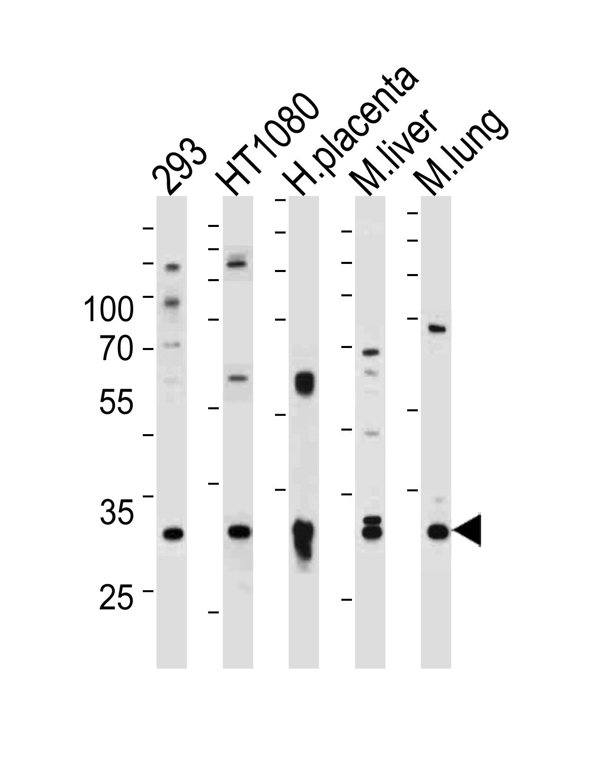
Western blot analysis of lysates from 293,HT1080 cell line,human placenta,mouse liver and lung tissue (from left to right),using SAR1A Antibody (Center)(Cat. #P32565).P32565 was diluted at 1:1000 at each lane. A goat anti-rabbit IgG H&L(HRP) at 1:5000 dilution was used as the secondary antibody.Lysates at 35ug per lane.

-
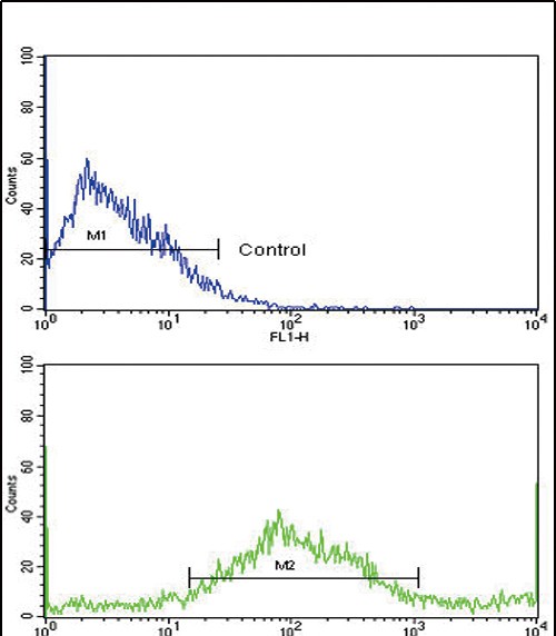
Flow cytometric analysis of NCI-H292 cells using SAR1A Antibody (Center)(bottom histogram) compared to a negative control cell (top histogram). FITC-conjugated goat-anti-rabbit secondary antibodies were used for the analysis.

-
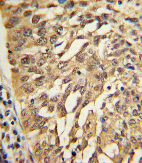
Formalin-fixed and paraffin-embedded human lung carcinoma with SAR1A Antibody (Center), which was peroxidase-conjugated to the secondary antibody, followed by DAB staining. This data demonstrates the use of this antibody for immunohistochemistry; clinical relevance has not been evaluated.


 鄂公网安备42018502007531号

