
Rabbit Polyclonal Antibody to EEF1B2
货号:
P32608
别名:
Elongation factor 1-beta, EF-1-beta, EEF1B2, EEF1B, EF1B
应用:
WB,IHC,IF,FCM
反应种属:
Human
抗体类型:
Primary antibody
Swissprot:
P24534
规格:
目录价
在线咨询
Description |
|---|
EF-1-beta and EF-1-delta stimulate the exchange of GDP bound to EF-1-alpha to GTP. |
Specification |
|
|---|---|
| Aliases | Elongation factor 1-beta, EF-1-beta, EEF1B2, EEF1B, EF1B |
| Entrez GeneID | 1933 |
| Swissprot | P24534 |
| WB Predicted band size | 24.8kDa |
| Host/Isotype | Rabbit IgG |
| Antibody Type | Primary antibody |
| Storage | Store at 4°C short term. Aliquot and store at -20°C long term. Avoid freeze/thaw cycles. |
| Species Reactivity | Human |
| Immunogen | This EEF1B2 antibody is generated from a rabbit immunized with a KLH conjugated synthetic peptide between 54-86 amino acids from the Central region of human EEF1B2. |
Application |
|
|---|---|
| WB | 1/1000 |
| IHC | 1/100-1/500 |
| IF | 1/25 |
| FCM | 1/25 |
Product Image
-
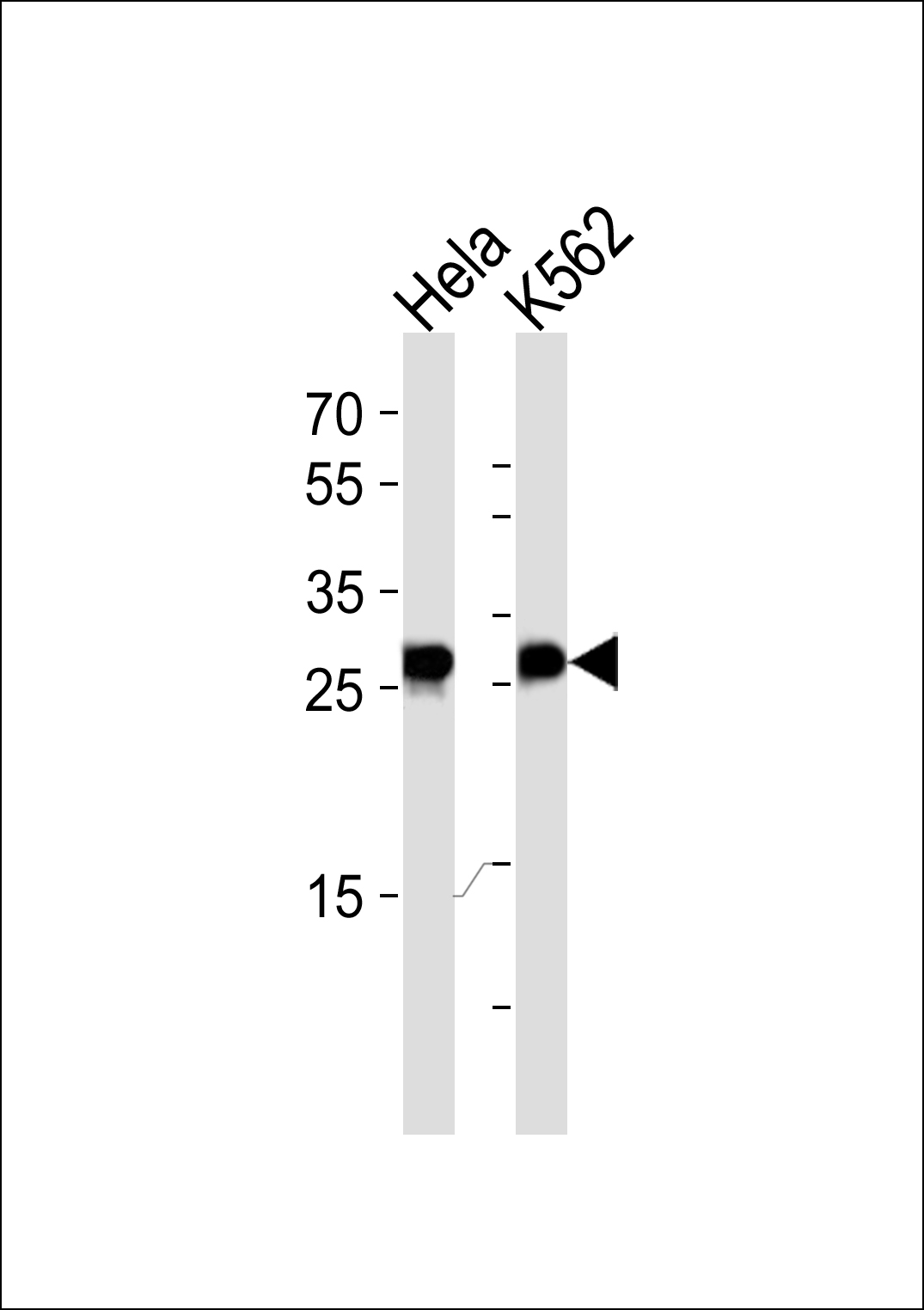
Western blot analysis of lysates from Hela, K562 cell line (from left to right), using EEF1B2 Antibody (Center)(Cat. #P32608). P32608 was diluted at 1:1000 at each lane. A goat anti-rabbit IgG H&L(HRP) at 1:5000 dilution was used as the secondary antibody. Lysates at 35ug per lane.

-
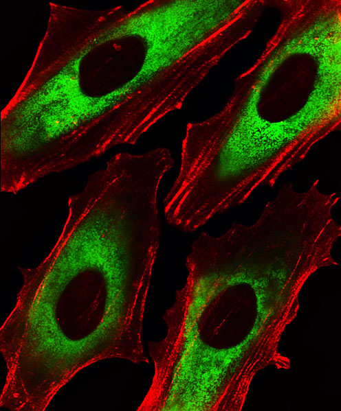
Fluorescent image of Hela cells stained with EEF1B2 Antibody (Center)(Cat#P32608). P32608 was diluted at 1:25 dilution. An Alexa Fluor 488-conjugated goat anti-rabbit lgG at 1:400 dilution was used as the secondary antibody (green). Cytoplasmic actin was counterstained with Alexa Fluor® 555 conjugated with Phalloidin (red).

-
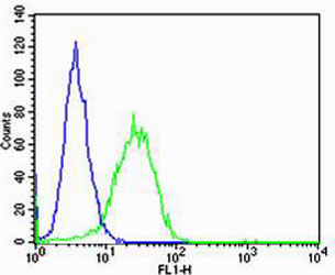
Flow cytometric analysis of Hela cells using EEF1B2 Antibody (Center)(green, Cat#P32608) compared to an isotype control of rabbit IgG(blue). P32608 was diluted at 1:25 dilution. An Alexa Fluor® 488 goat anti-rabbit lgG at 1:400 dilution was used as the secondary antibody.

-
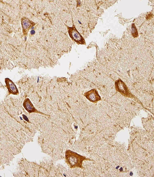
Immunohistochemical analysis of paraffin-embedded H. brain section using EEF1B2 Antibody (Center)(Cat#P32608). P32608 was diluted at 1:25 dilution. A peroxidase-conjugated goat anti-rabbit IgG at 1:400 dilution was used as the secondary antibody, followed by DAB staining.


 鄂公网安备42018502007531号

