
Mouse Monoclonal Antibody to CAPN2
货号:
P32636
别名:
Calpain-2 catalytic subunit, Calcium-activated neutral proteinase 2, CANP 2, Calpain M-type, Calpain large polypeptide L2, Calpain-2 large subunit, Millimolar-calpain, M-calpain, CAPN2, CANPL2
应用:
WB,IHC,FCM
反应种属:
Human, Mouse
抗体类型:
Primary antibody
Swissprot:
P17655
规格:
目录价
在线咨询
Description |
|---|
Calcium-regulated non-lysosomal thiol-protease which catalyze limited proteolysis of substrates involved in cytoskeletal remodeling and signal transduction. |
Specification |
|
|---|---|
| Aliases | Calpain-2 catalytic subunit, Calcium-activated neutral proteinase 2, CANP 2, Calpain M-type, Calpain large polypeptide L2, Calpain-2 large subunit, Millimolar-calpain, M-calpain, CAPN2, CANPL2 |
| Entrez GeneID | 824 |
| Swissprot | P17655 |
| WB Predicted band size | 80.0kDa |
| Host/Isotype | Mouse IgG2b |
| Antibody Type | Primary antibody |
| Storage | Store at 4°C short term. Aliquot and store at -20°C long term. Avoid freeze/thaw cycles. |
| Species Reactivity | Human, Mouse |
| Immunogen | This CAPN2 antibody is generated from a mouse immunized with a KLH conjugated synthetic peptide between amino acids from the human region of human CAPN2. |
Application |
|
|---|---|
| WB | 1/1000 |
| IHC | 1/100-1/500 |
| FCM | 1/100 |
Product Image
-
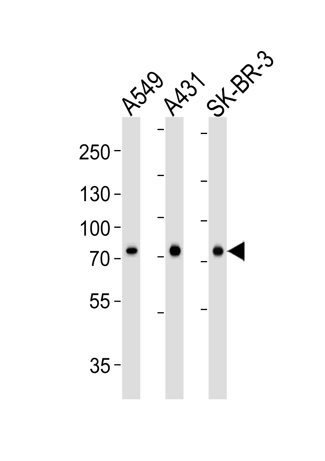
Western blot analysis of lysates from A549, A431, SK-BR-3 cell line (from left to right), using CAPN2 Antibody(Cat. #P32636). P32636 was diluted at 1:1000 at each lane. A goat anti-mouse IgG H&L(HRP) at 1:3000 dilution was used as the secondary antibody. Lysates at 35μg per lane.

-
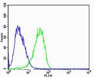
Flow cytometric analysis of U-87 MG cells using CAPN2 Antibody(green, Cat#P32636) compared to an isotype control of mouse IgG2b(blue). AP20600c was diluted at 1:100 dilution. An Alexa Fluor® 488 goat anti-mouse lgG at 1:400 dilution was used as the secondary antibody.

-
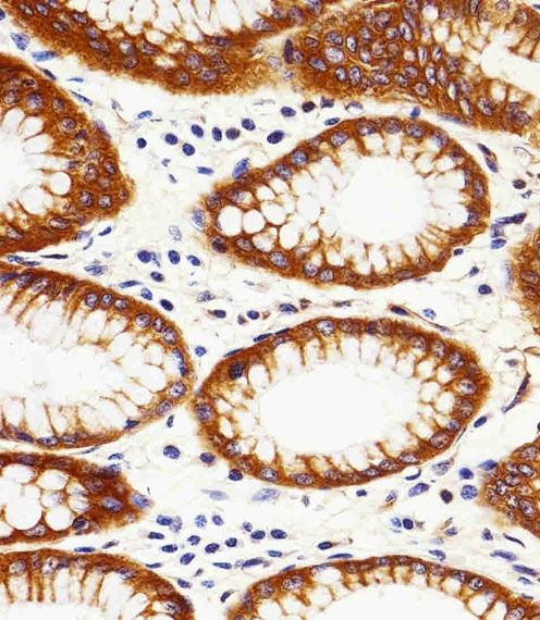
Immunohistochemical analysis of paraffin-embedded H.stomach section using CAPN2 Antibody(Cat#P32636). P32636 was diluted at 1:25 dilution. A peroxidase-conjugated goat anti-mouse IgG at 1:400 dilution was used as the secondary antibody, followed by DAB staining.


 鄂公网安备42018502007531号

