
Rabbit Polyclonal Antibody to TFAM
货号:
P32879
别名:
Transcription factor A, mitochondrial, mtTFA, Mitochondrial transcription factor 1, MtTF1, Transcription factor 6, TCF-6, Transcription factor 6-like 2, TFAM, TCF6, TCF6L2
应用:
WB,IHC,IF,FCM
反应种属:
Human
抗体类型:
Primary antibody
Swissprot:
Q00059
规格:
目录价
在线咨询
Description |
|---|
This gene encodes a mitochondrial transcription factor that is a key activator of mitochondrial transcription as well as a participant in mitochondrial genome replication. Studies in mice have demonstrated that this gene product is required to regulate the mitochondrial genome copy number and is essential for embryonic development. A mouse model for Kearns-Sayre syndrome was produced when expression of this gene was eliminated by targeted disruption in heart and muscle cells. |
Specification |
|
|---|---|
| Aliases | Transcription factor A, mitochondrial, mtTFA, Mitochondrial transcription factor 1, MtTF1, Transcription factor 6, TCF-6, Transcription factor 6-like 2, TFAM, TCF6, TCF6L2 |
| Entrez GeneID | 7019 |
| Swissprot | Q00059 |
| WB Predicted band size | 29.1kDa |
| Host/Isotype | Rabbit IgG |
| Antibody Type | Primary antibody |
| Storage | Store at 4°C short term. Aliquot and store at -20°C long term. Avoid freeze/thaw cycles. |
| Species Reactivity | Human |
| Immunogen | This TFAM antibody is generated from rabbits immunized with a KLH conjugated synthetic peptide between 216-246 amino acids from the C-terminal region of human TFAM. |
| Formulation | Purified antibody in PBS with 0.05% sodium azide. |
Application |
|
|---|---|
| WB | 1/1000 |
| IHC | 1/100-1/500 |
| IF | 1/10-1/50 |
| FCM | 1/10-1/50 |
Product Image
-
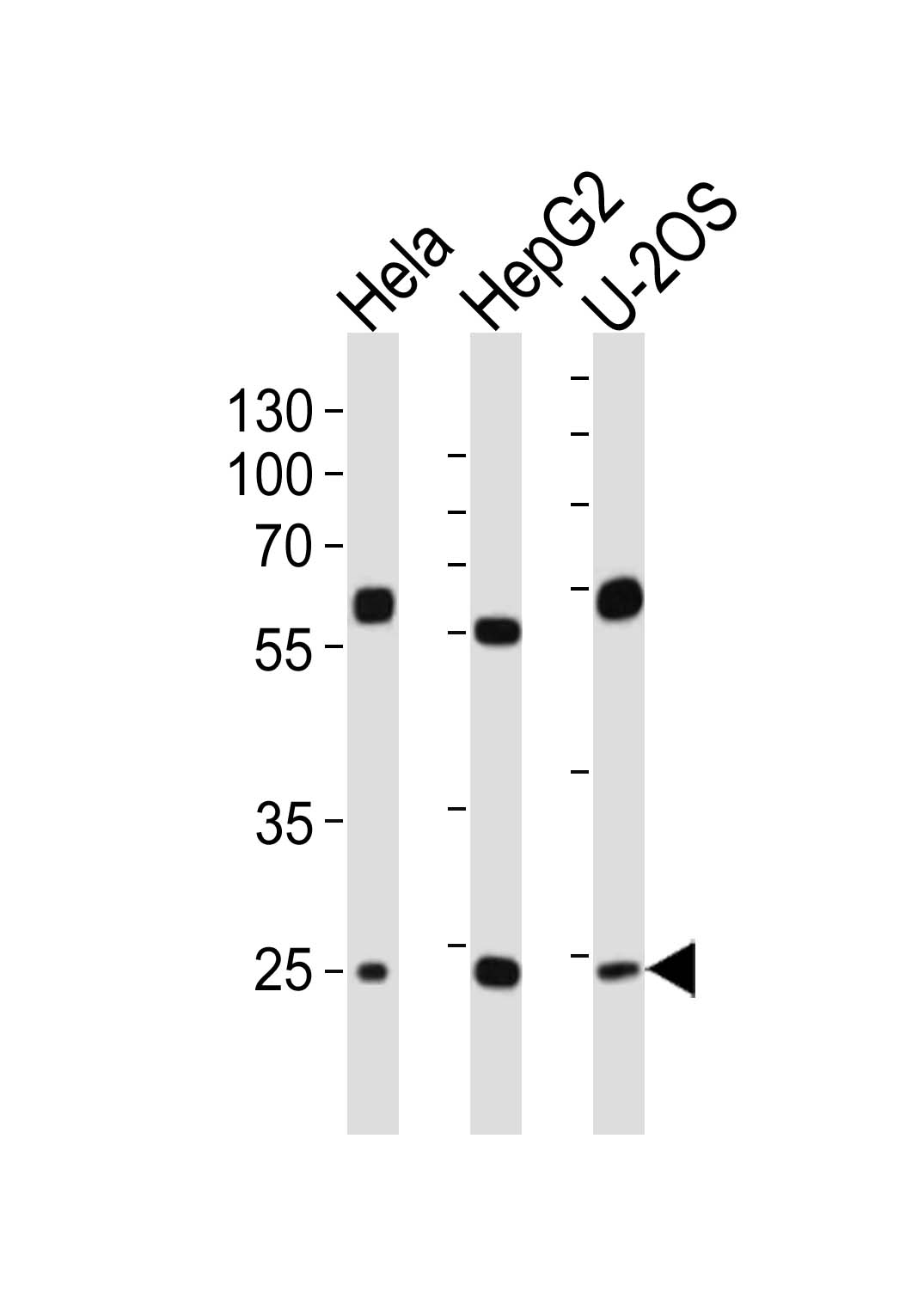
Western blot analysis of lysates from Hela, HepG2, U-2OS cell line (from left to right), using TFAM Antibody (C-term)(Cat. #P32879). P32879 was diluted at 1:1000 at each lane. A goat anti-rabbit IgG H&L(HRP) at 1:10000 dilution was used as the secondary antibody. Lysates at 20ug per lane.

-
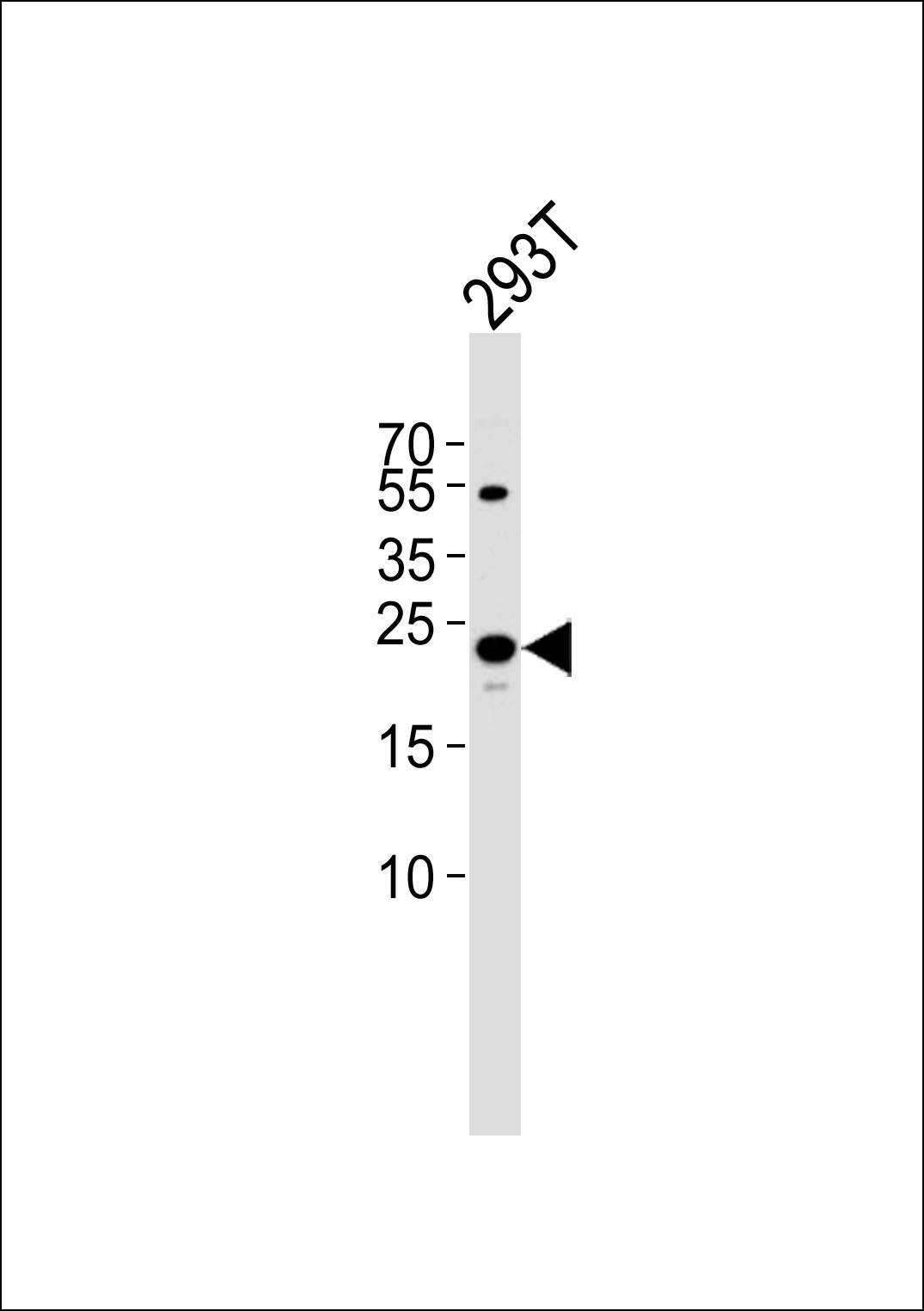
Western blot analysis of lysate from 293T cell line, using TFAM Antibody (C-term) (Cat. #P32879). P32879 was diluted at 1:1000. A goat anti-rabbit IgG H&L(HRP) at 1:5000 dilution was used as the secondary antibody. Lysate at 35ug.

-
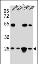
TFAM Antibody (C-term) (Cat. #P32879) western blot analysis in Hela,Jurkat,K562,MCF-7 cell line lysates (35ug/lane).This demonstrates the TFAM antibody detected the TFAM protein (arrow).

-
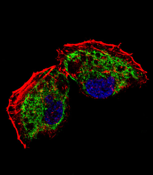
Fluorescent confocal image of NCI-H460 cell stained with TFAM Antibody (C-term)(Cat#P32879). NCI-H460 cells were fixed with 4% PFA (20 min), permeabilized with Triton X-100 (0.1%, 10 min), then incubated with TFAM primary antibody (1:25, 1 h at 37℃). For secondary antibody, Alexa Fluor® 488 conjugated donkey anti-rabbit antibody (green) was used (1:400, 50 min at 37℃).Cytoplasmic actin was counterstained with Alexa Fluor® 555 (red) conjugated Phalloidin (7units/ml, 1 h at 37℃). Nuclei were counterstained with DAPI (blue) (10 µg/ml, 10 min).TFAM immunoreactivity is localized to mitochondrion significantly.

-
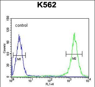
TFAM Antibody (C-term) (Cat. #P32879) flow cytometric analysis of K562 cells (right histogram) compared to a negative control cell (left histogram).FITC-conjugated goat-anti-rabbit secondary antibodies were used for the analysis.

-
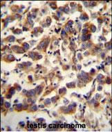
TFAM antibody (C-term) (Cat. #P32879) immunohistochemistry analysis in formalin fixed and paraffin embedded human testis carcinoma followed by peroxidase conjugation of the secondary antibody and DAB staining. This data demonstrates the use of the TFAM antibody (C-term) for immunohistochemistry. Clinical relevance has not been evaluated.


 鄂公网安备42018502007531号

