
Mouse Monoclonal Antibody to ALDH6A1
货号:
P32976
别名:
Methylmalonate-semialdehyde dehydrogenase [acylating], mitochondrial, MMSDH, Malonate-semialdehyde dehydrogenase [acylating], Aldehyde dehydrogenase family 6 member A1, ALDH6A1, MMSDH
应用:
WB,IHC,IF
反应种属:
Human, Mouse
抗体类型:
Primary antibody
Swissprot:
Q02252
规格:
目录价
在线咨询
Description |
|---|
This protein belongs to the aldehyde dehydrogenases family of proteins. This enzyme plays a role in the valine and pyrimidine catabolic pathways. The product of this gene, a mitochondrial methylmalonate semialdehyde dehydrogenase, catalyzes the irreversible oxidative decarboxylation of malonate and methylmalonate semialdehydes to acetyl- and propionyl-CoA. Methylmalonate semialdehyde dehydrogenase deficiency is characterized by elevated beta-alanine, 3-hydroxypropionic acid, and both isomers of 3-amino and 3-hydroxyisobutyric acids in urine organic acids. |
Specification |
|
|---|---|
| Aliases | Methylmalonate-semialdehyde dehydrogenase [acylating], mitochondrial, MMSDH, Malonate-semialdehyde dehydrogenase [acylating], Aldehyde dehydrogenase family 6 member A1, ALDH6A1, MMSDH |
| Entrez GeneID | 57840 |
| Swissprot | Q02252 |
| WB Predicted band size | 57.8kDa |
| Host/Isotype | Mouse IgG1 |
| Antibody Type | Primary antibody |
| Storage | Store at 4°C short term. Aliquot and store at -20°C long term. Avoid freeze/thaw cycles. |
| Species Reactivity | Human, Mouse |
| Immunogen | This ALDH6A1 antibody is generated from mouse immunized with ALDH6A1 recombinant protein. |
| Formulation | Purified antibody in TBS with 0.05% sodium azide. |
Application |
|
|---|---|
| WB | 1/1000 |
| IHC | 1/100-1/500 |
| IF | 1/25 |
Product Image
-
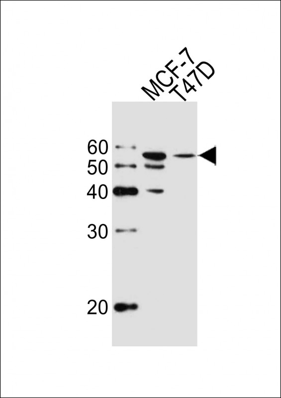
Western blot analysis of lysates from MCF-7, T47D cell line (from left to right), using ALDH6A1 Antibody(Cat. #P32976). P32976 was diluted at 1:1000 at each lane. A goat anti-mouse IgG H&L(HRP) at 1:10000 dilution was used as the secondary antibody. Lysates at 20μg per lane.

-
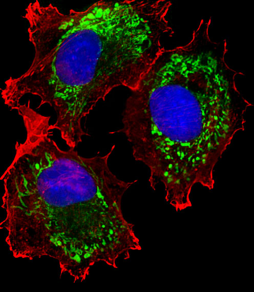
Fluorescent image of MCF-7 cells stained with ALDH6A1 Antibody (Cat#P32976). P32976 was diluted at 1:25 dilution. An Alexa Fluor® 488-conjugated goat anti-mouse lgG at 1:400 dilution was used as the secondary antibody (green). DAPI was used to stain the cell nuclear (blue). Cytoplasmic actin was counterstained with Alexa Fluor® 555 conjugated with Phalloidin (red).

-
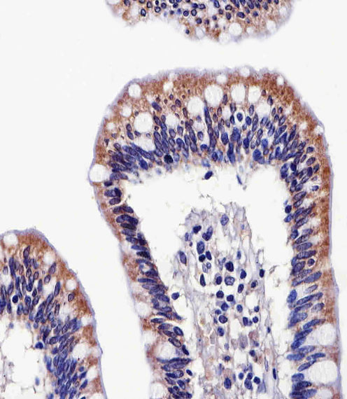
Immunohistochemical analysis of paraffin-embedded H.colon section using ALDH6A1 Antibody(Cat#P32976). P32976 was diluted at 1:25 dilution. A peroxidase-conjugated goat anti-mouse IgG at 1:400 dilution was used as the secondary antibody, followed by DAB staining.

-
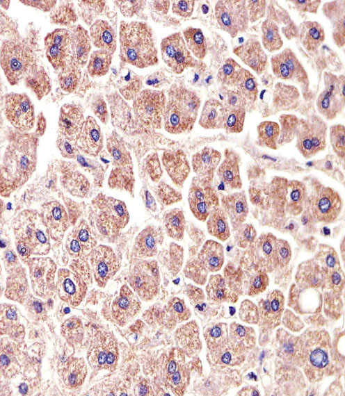
Immunohistochemical analysis of paraffin-embedded H.liver section using ALDH6A1 Antibody(Cat#P32976). P32976 was diluted at 1:25 dilution. A peroxidase-conjugated goat anti-mouse IgG at 1:400 dilution was used as the secondary antibody, followed by DAB staining.


 鄂公网安备42018502007531号

