
Rabbit Polyclonal Antibody to PAX4
货号:
P33123
别名:
Paired box protein Pax-4, PAX4
应用:
WB,IHC,FCM
反应种属:
Human, Mouse, Rat
抗体类型:
Primary antibody
Swissprot:
O43316
规格:
目录价
在线咨询
Description |
|---|
PAX4 is a member of the paired box (PAX) family of transcription factors. These proteins play critical roles during fetal development and cancer growth. The paired box 4 gene is involved in pancreatic islet development and mouse studies have demonstrated a role for this gene in differentiation of insulin-producing beta cells. |
Specification |
|
|---|---|
| Aliases | Paired box protein Pax-4, PAX4 |
| Swissprot | O43316 |
| WB Predicted band size | 37.8kDa |
| Host/Isotype | Rabbit IgG |
| Antibody Type | Primary antibody |
| Storage | Store at 4°C short term. Aliquot and store at -20°C long term. Avoid freeze/thaw cycles. |
| Species Reactivity | Human, Mouse, Rat |
| Immunogen | This PAX4 antibody is generated from rabbits immunized with a KLH conjugated synthetic peptide between 171-200 amino acids from the Central region of human PAX4. |
| Formulation | Purified antibody in PBS with 0.05% sodium azide,1%BSA and 50% glycerol.prepared by Saturated Ammonium Sulfate (SAS) . |
Application |
|
|---|---|
| WB | 1/1000-1/2000 |
| IHC | 1/100-1/500 |
| FCM | 1/10-1/50 |
Product Image
-
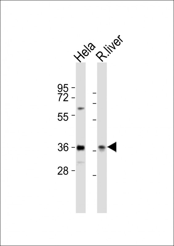
All lanes : Anti-PAX4 Antibody (Center) at 1:2000 dilution Lane 1: Hela whole cell lysates Lane 2: rat liver lysates Lysates/proteins at 20 µg per lane. Secondary Goat Anti-Rabbit IgG, (H+L), Peroxidase conjugated at 1/10000 dilution Predicted band size : 38 kDa Blocking/Dilution buffer: 5% NFDM/TBST.

-
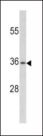
Western blot analysis of PAX4 antibody (Center) (RB20216) in CEM cell line lysates (35ug/lane). PAX4 (arrow) was detected using the purified Pab.

-
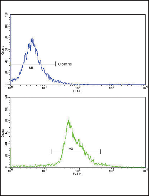
Flow cytometric analysis of widr cells using PAX4 Antibody (Center)(bottom histogram) compared to a negative control cell (top histogram)FITC-conjugated goat-anti-rabbit secondary antibodies were used for the analysis.

-
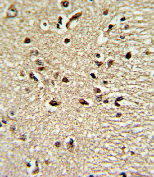
Formalin-fixed and paraffin-embedded human brain tissue reacted with PAX4 Antibody (Center), which was peroxidase-conjugated to the secondary antibody, followed by DAB staining. This data demonstrates the use of this antibody for immunohistochemistry; clinical relevance has not been evaluated.


 鄂公网安备42018502007531号

