
Mouse Monoclonal Antibody to HCK
货号:
P33212
别名:
Tyrosine-protein kinase HCK, Hematopoietic cell kinase, Hemopoietic cell kinase, p59-HCK/p60-HCK, p59Hck, p61Hck, HCK
应用:
WB,IF
反应种属:
Human, Mouse, Rat
抗体类型:
Primary antibody
Swissprot:
P08631
规格:
目录价
在线咨询
Description |
|---|
Non-receptor tyrosine-protein kinase found in hematopoietic cells that transmits signals from cell surface receptors and plays an important role in the regulation of innate immune responses, including neutrophil, monocyte, macrophage and mast cell functions, phagocytosis, cell survival and proliferation, cell adhesion and migration. Acts downstream of receptors that bind the Fc region of immunoglobulins, such as FCGR1A and FCGR2A, but also CSF3R, PLAUR, the receptors for IFNG, IL2, IL6 and IL8, and integrins, such as ITGB1 and ITGB2. During the phagocytic process, mediates mobilization of secretory lysosomes, degranulation, and activation of NADPH oxidase to bring about the respiratory burst. Plays a role in the release of inflammatory molecules. Promotes reorganization of the actin cytoskeleton and actin polymerization, formation of podosomes and cell protrusions. Inhibits TP73-mediated transcription activation and TP73-mediated apoptosis. Phosphorylates CBL in response to activation of immunoglobulin gamma Fc region receptors. Phosphorylates ADAM15, BCR, ELMO1, FCGR2A, GAB1, GAB2, RAPGEF1, STAT5B, TP73, VAV1 and WAS. |
Specification |
|
|---|---|
| Aliases | Tyrosine-protein kinase HCK, Hematopoietic cell kinase, Hemopoietic cell kinase, p59-HCK/p60-HCK, p59Hck, p61Hck, HCK |
| Entrez GeneID | 3055 |
| Swissprot | P08631 |
| WB Predicted band size | 59.6kDa |
| Host/Isotype | Mouse IgG1 |
| Antibody Type | Primary antibody |
| Storage | Store at 4°C short term. Aliquot and store at -20°C long term. Avoid freeze/thaw cycles. |
| Species Reactivity | Human, Mouse, Rat |
| Immunogen | This HCK antibody is generated from a mouse immunized with a recombinant protein of human HCK. |
Application |
|
|---|---|
| WB | 1/2002-1/4000 |
| IF | 1/25 |
Product Image
-
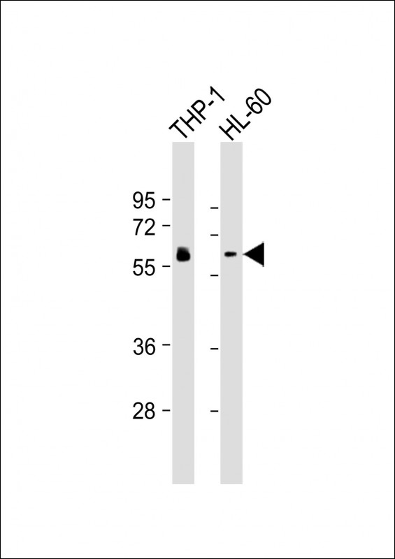
All lanes : Anti-HCK Antibody at 1:2000-1:4000 dilution Lane 1: THP-1 whole cell lysates Lane 2: HL-60 whole cell lysates Lysates/proteins at 20 μg per lane. Secondary Goat Anti-mouse IgG, (H+L), Peroxidase conjugated at 1/10000 dilution. Predicted band size : 60 kDa Blocking/Dilution buffer: 5% NFDM/TBST.

-
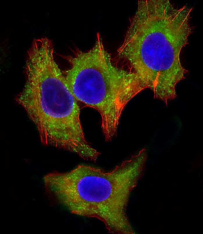
Immunofluorescent analysis of 4% paraformaldehyde-fixed, 0.1% Triton X-100 permeabilized HepG2 (human liver hepatocellular carcinoma cell line) cells labeling HCK with P33212 at 1/25 dilution, followed by Dylight® 488-conjugated goat anti-mouse IgG secondary antibody at 1/200 dilution (green). Immunofluorescence image showing cytoplasm staining on HepG2 cell line. Cytoplasmic actin is detected with Dylight® 554 Phalloidin at 1/100 dilution (red).The nuclear counter stain is DAPI (blue).


 鄂公网安备42018502007531号

