
Rabbit Polyclonal Antibody to RNF7 (N-term)
货号:
P33258
别名:
RING-box protein 2, Rbx2, CKII beta-binding protein 1, CKBBP1, RING finger protein 7, Regulator of cullins 2, Sensitive to apoptosis gene protein, RNF7, RBX2, ROC2, SAG
应用:
WB,IHC
反应种属:
Human, Mouse
抗体类型:
Primary antibody
Swissprot:
Q9UBF6
规格:
目录价
在线咨询
Description |
|---|
Probable component of the SCF (SKP1-CUL1-F-box protein) E3 ubiquitin ligase complex which mediates the ubiquitination and subsequent proteasomal degradation of target proteins involved in cell cycle progression, signal transduction and transcription. Through the RING-type zinc finger, seems to recruit the E2 ubiquitination enzyme to the complex and brings it into close proximity to the substrate. Promotes the neddylation of CUL5 via its interaction with UBE2F. May play a role in protecting cells from apoptosis induced by redox agents. |
Specification |
|
|---|---|
| Aliases | RING-box protein 2, Rbx2, CKII beta-binding protein 1, CKBBP1, RING finger protein 7, Regulator of cullins 2, Sensitive to apoptosis gene protein, RNF7, RBX2, ROC2, SAG |
| Entrez GeneID | 9616 |
| Swissprot | Q9UBF6 |
| WB Predicted band size | 12.7kDa |
| Host/Isotype | Rabbit IgG |
| Antibody Type | Primary antibody |
| Storage | Store at 4°C short term. Aliquot and store at -20°C long term. Avoid freeze/thaw cycles. |
| Species Reactivity | Human, Mouse |
| Immunogen | This RNF7 antibody is generated from a rabbit immunized with a KLH conjugated synthetic peptide between 25-57 amino acids from the N-terminal region of human RNF7. |
Application |
|
|---|---|
| WB | 1/8000 |
| IHC | 1/100-1/500 |
Product Image
-
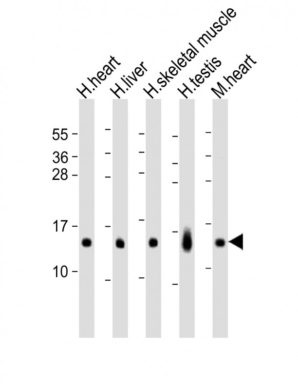
All lanes : Anti-RNF7 Antibody (N-term) at 1:8000 dilution Lane 1: human heart lysates Lane 2: human liver lysates Lane 3: human skeletal muscle lysates Lane 4: human testis lysates Lane 5: mouse heart lysates Lysates/proteins at 20 µg per lane. Secondary Goat Anti-Rabbit IgG, (H+L), Peroxidase conjugated at 1/10000 dilution. Predicted band size : 13 kDa Blocking/Dilution buffer: 5% NFDM/TBST.

-
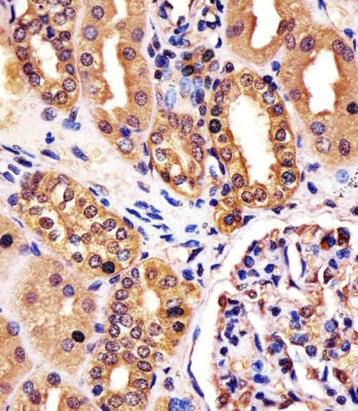
P33258 staining RNF7 in human kidney tissue sections by Immunohistochemistry (IHC-P - paraformaldehyde-fixed, paraffin-embedded sections). Tissue was fixed with formaldehyde and blocked with 3% BSA for 0. 5 hour at room temperature; antigen retrieval was by heat mediation with a citrate buffer (pH6). Samples were incubated with primary antibody (1/25) for 1 hours at 37°C. A undiluted biotinylated goat polyvalent antibody was used as the secondary antibody.


 鄂公网安备42018502007531号

