
Rabbit Polyclonal Antibody to TBK1 (S172)
货号:
P33668
别名:
Serine/threonine-protein kinase TBK1, NF-kappa-B-activating kinase, T2K, TANK-binding kinase 1, TBK1, NAK
应用:
WB,IHC
反应种属:
Human, Mouse
抗体类型:
Primary antibody
Swissprot:
Q9UHD2
规格:
目录价
在线咨询
Description |
|---|
The NF-kappa-B (NFKB) complex of proteins is inhibited by I-kappa-B (IKB) proteins, which inactivate NFKB by trapping it in the cytoplasm. Phosphorylation of serine residues on the IKB proteins by IKB kinases marks them for destruction via the ubiquitination pathway, thereby allowing activation and nuclear translocation of the NFKB complex. TKB is similar to IKB kinases and can mediate NFKB activation in response to certain growth factors. The protein can form a complex with the IKB protein TANK and TRAF2 and release the NFKB inhibition caused by TANK. |
Specification |
|
|---|---|
| Aliases | Serine/threonine-protein kinase TBK1, NF-kappa-B-activating kinase, T2K, TANK-binding kinase 1, TBK1, NAK |
| Entrez GeneID | 29110 |
| Swissprot | Q9UHD2 |
| WB Predicted band size | 83.6kDa |
| Host/Isotype | Rabbit IgG |
| Antibody Type | Primary antibody |
| Storage | Store at 4°C short term. Aliquot and store at -20°C long term. Avoid freeze/thaw cycles. |
| Species Reactivity | Human, Mouse |
| Immunogen | This TBK antibody is generated from rabbits immunized with a KLH conjugated synthetic peptide between 150-181 amino acids from human TBK. |
| Formulation | Purified antibody in PBS with 0.05% sodium azide. |
Application |
|
|---|---|
| WB | 1/2000 |
| IHC | 1/100-1/500 |
Product Image
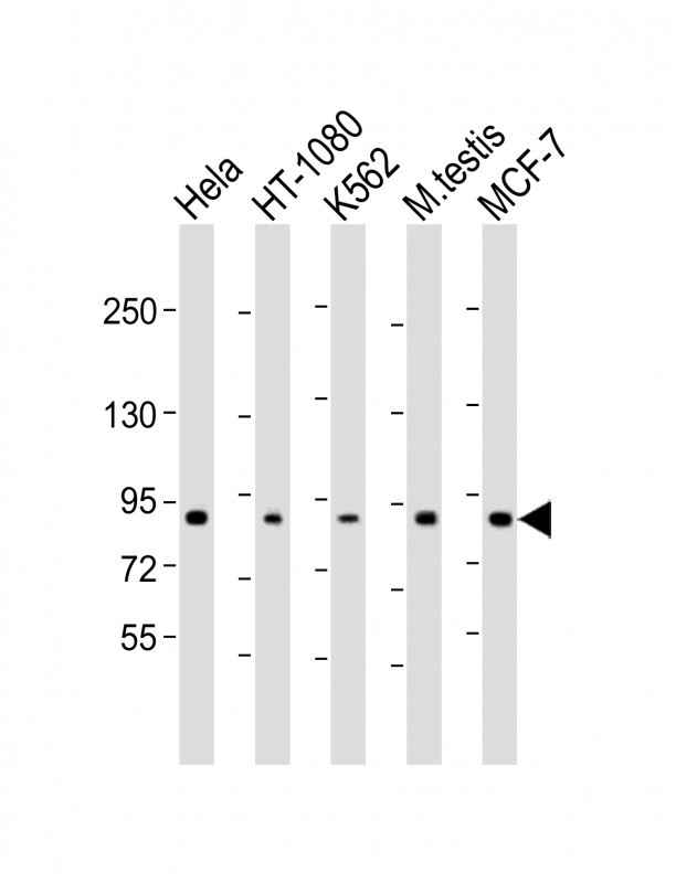
-
All lanes : Anti-TBK1 Antibody (S172) at 1:2000 dilution Lane 1: Hela whole cell lysate Lane 2: HT-1080 whole cell lysate Lane 3: K562 whole cell lysate Lane 4: mouse testis lysate Lane 5: MCF-7 whole cell lysate Lysates/proteins at 20 µg per lane. Secondary Goat Anti-Rabbit IgG, (H+L), Peroxidase conjugated at 1/10000 dilution. Predicted band size :83kDa Blocking/Dilution buffer: 5% NFDM/TBST.

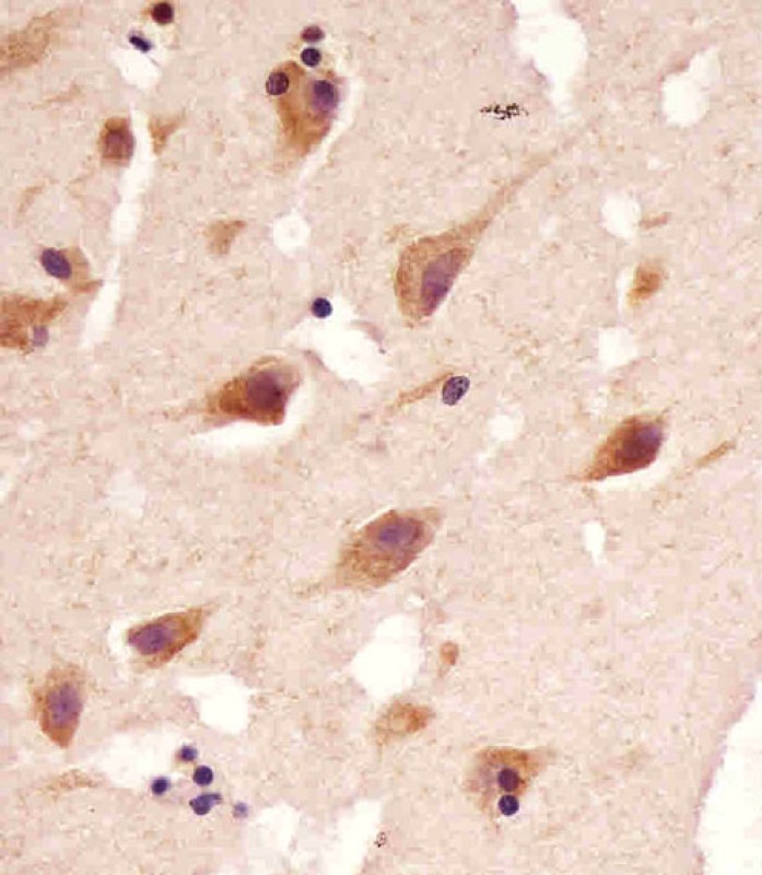
-
P33668 staining TBK1 in human brain tissue sections by Immunohistochemistry (IHC-P - paraformaldehyde-fixed, paraffin-embedded sections). Tissue was fixed with formaldehyde and blocked with 3% BSA for 0. 5 hour at room temperature; antigen retrieval was by heat mediation with a citrate buffer (pH6). Samples were incubated with primary antibody (1/25) for 1 hours at 37°C. A undiluted biotinylated goat polyvalent antibody was used as the secondary antibody.


 鄂公网安备42018502007531号

