
Rabbit Polyclonal Antibody to HSD17B3
货号:
P34051
别名:
Testosterone 17-beta-dehydrogenase 3, 17-beta-hydroxysteroid dehydrogenase type 3, 17-beta-HSD 3, Testicular 17-beta-hydroxysteroid dehydrogenase, HSD17B3, EDH17B3
应用:
WB,IHC,FCM
反应种属:
Human
抗体类型:
Primary antibody
Swissprot:
P37058
规格:
目录价
在线咨询
Description |
|---|
This isoform of 17 beta-hydroxysteroid dehydrogenase is expressed predominantly in the testis and catalyzes the conversion of androstenedione to testosterone. It preferentially uses NADP as cofactor. Deficiency can result in male pseudohermaphroditism with gynecomastia. |
Specification |
|
|---|---|
| Aliases | Testosterone 17-beta-dehydrogenase 3, 17-beta-hydroxysteroid dehydrogenase type 3, 17-beta-HSD 3, Testicular 17-beta-hydroxysteroid dehydrogenase, HSD17B3, EDH17B3 |
| Entrez GeneID | 3293 |
| Swissprot | P37058 |
| WB Predicted band size | 34.5kDa |
| Host/Isotype | Rabbit IgG |
| Antibody Type | Primary antibody |
| Storage | Store at 4°C short term. Aliquot and store at -20°C long term. Avoid freeze/thaw cycles. |
| Species Reactivity | Human |
| Immunogen | This HSD17B3 antibody is generated from rabbits immunized with a KLH conjugated synthetic peptide between 89-118 amino acids from the Central region of human HSD17B3. |
| Formulation | Purified antibody in PBS with 0.05% sodium azide. |
Application |
|
|---|---|
| WB | 1/1000 |
| IHC | 1/100-1/500 |
| FCM | 1/10-1/50 |
Product Image
-
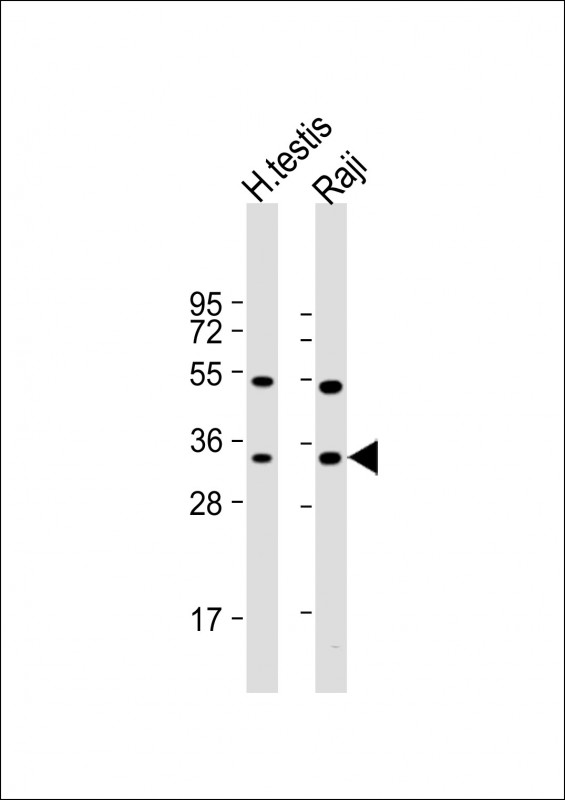
All lanes : Anti-HSD17B3 Antibody (Center) at 1:1000 dilution Lane 1: human testis lysate Lane 2: Raji whole cell lysate Lysates/proteins at 20 µg per lane. Secondary Goat Anti-Rabbit IgG, (H+L), Peroxidase conjugated at 1/10000 dilution. Predicted band size : 35 kDa Blocking/Dilution buffer: 5% NFDM/TBST.

-
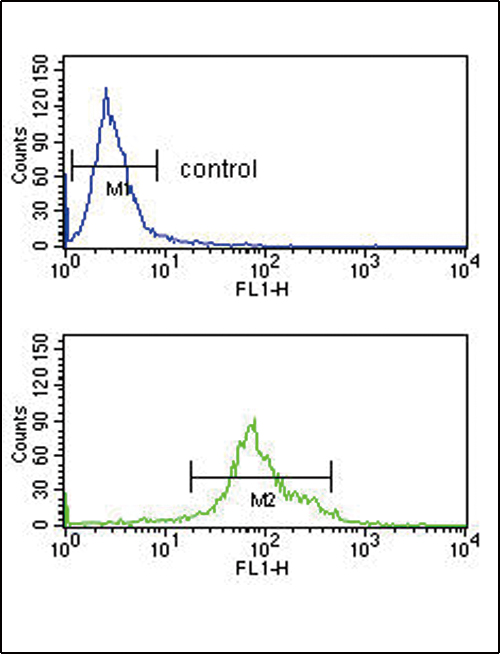
HSD17B3 Antibody (Center) (Cat. #P34051) flow cytometry analysis of K562 cells (bottom histogram) compared to a negative control cell (top histogram).FITC-conjugated goat-anti-rabbit secondary antibodies were used for the analysis.

-
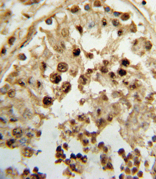
Formalin-fixed and paraffin-embedded human testis tissue reacted with HSD17B3 Antibody (Center), which was peroxidase-conjugated to the secondary antibody, followed by DAB staining. This data demonstrates the use of this antibody for immunohistochemistry; clinical relevance has not been evaluated.


 鄂公网安备42018502007531号

