
Rabbit Polyclonal Antibody to NANOG (S285)
货号:
P34110
别名:
Homeobox protein NANOG, Homeobox transcription factor Nanog, hNanog, NANOG
应用:
WB,FCM
反应种属:
Human
抗体类型:
Primary antibody
Swissprot:
Q9H9S0
规格:
目录价
在线咨询
Description |
|---|
NANOG is a Ttranscription regulator involved in inner cell mass and embryonic stem (ES) cels proliferation and self-renewal. It imposes pluripotency on ES cells and prevents their differentiation towards extraembryonic endoderm and trophectoderm lineages. This protein blocks bone morphogenetic protein-induced mesoderm differentiation of ES cells by physically interacting with SMAD1 and interfering with the recruitment of coactivators to the active SMAD transcriptional complexes. NANOG acts as a transcriptional activator or repressor. It binds optimally to the DNA consensus sequence 5'-[CG][GA][CG]C[GC]ATTAN[GC]-3'. When overexpressed, this protein promotes cells to enter into S phase and proliferation. |
Specification |
|
|---|---|
| Aliases | Homeobox protein NANOG, Homeobox transcription factor Nanog, hNanog, NANOG |
| Entrez GeneID | 79923 |
| Swissprot | Q9H9S0 |
| WB Predicted band size | 34.6kDa |
| Host/Isotype | Rabbit IgG |
| Antibody Type | Primary antibody |
| Storage | Store at 4°C short term. Aliquot and store at -20°C long term. Avoid freeze/thaw cycles. |
| Species Reactivity | Human |
| Immunogen | This NANOG antibody is generated from rabbits immunized with a KLH conjugated synthetic peptide between 267-292 amino acids from human NANOG. |
| Formulation | Purified antibody in PBS with 0.05% sodium azide. |
Application |
|
|---|---|
| WB | 1/1000 |
| FCM | 1/10-1/50 |
Product Image
-
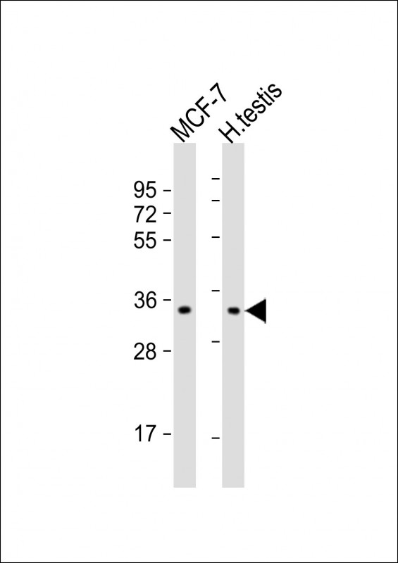
All lanes : Anti-NANOG Antibody (S285) at 1:1000 dilution Lane 1: MCF-7 whole cell lysate Lane 2: human testis lysate Lysates/proteins at 20 µg per lane. Secondary Goat Anti-Rabbit IgG, (H+L), Peroxidase conjugated at 1/10000 dilution. Predicted band size : 35 kDa Blocking/Dilution buffer: 5% NFDM/TBST.

-
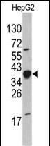
Western blot analysis of NANOG Antibody (S285) (Cat.# AP1486e) in HepG2 cell line lysates (35ug/lane). NANOG (arrow) was detected using the purified Pab.

-
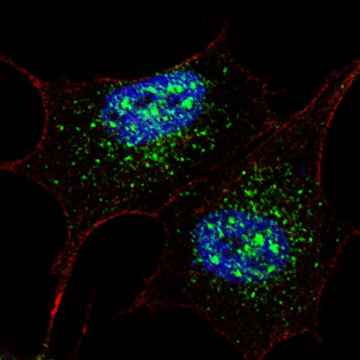
Fluorescent confocal image of SY5Y cells stained with AP1486e NANOG (S285) antibody. SY5Y cells were fixed with 4% PFA (20 min), permeabilized with Triton X-100 (0.2%, 30 min), then incubated with AP1486e NANOG (S285) primary antibody (1:200, 2 h at room temperature). For secondary antibody, Alexa Fluor® 488 conjugated donkey anti-rabbit antibody (green) was used (1:1000, 1h). Cytoplasmic actin was counterstained with Alexa Fluor® 555 (red) conjugated Phalloidin (5.25 μM, 25 min). Nuclei were counterstained with Hoechst 33342 (blue) (10 µg/ml, 3 min). Nanog immunoreactivity is localized mainly to the nuclei and also to the cytoplasm.


 鄂公网安备42018502007531号

