
Rabbit Polyclonal Antibody to GIT1
货号:
P34211
别名:
ARF GTPase-activating protein GIT1, ARF GAP GIT1, Cool-associated and tyrosine-phosphorylated protein 1, CAT-1, CAT1, G protein-coupled receptor kinase-interactor 1, GRK-interacting protein 1, GIT1
应用:
WB,IHC
反应种属:
Human
抗体类型:
Primary antibody
Swissprot:
Q9Y2X7
规格:
目录价
在线咨询
Description |
|---|
GIT1 is a GTPase-activating protein for the ADP ribosylation factor family. It may serve as a scaffold to bring together molecules to form signaling modules controlling vesicle trafficking, adhesion and cytoskeletal organization. It increases the speed of cell migration, as well as the size and rate of formation of protrusions, possibly by targeting PAK1 to adhesions and the leading edge of lamellipodia. |
Specification |
|
|---|---|
| Aliases | ARF GTPase-activating protein GIT1, ARF GAP GIT1, Cool-associated and tyrosine-phosphorylated protein 1, CAT-1, CAT1, G protein-coupled receptor kinase-interactor 1, GRK-interacting protein 1, GIT1 |
| Entrez GeneID | 28964 |
| Swissprot | Q9Y2X7 |
| WB Predicted band size | 84.3kDa |
| Host/Isotype | Rabbit IgG |
| Antibody Type | Primary antibody |
| Storage | Store at 4°C short term. Aliquot and store at -20°C long term. Avoid freeze/thaw cycles. |
| Species Reactivity | Human |
| Immunogen | This GIT1 antibody is generated from rabbits immunized with a KLH conjugated synthetic peptide between 485-512 amino acids from the Central region of human GIT1. |
| Formulation | Purified antibody in PBS with 0.05% sodium azide,1%BSA and 50% glycerol.prepared by Saturated Ammonium Sulfate (SAS) . |
Application |
|
|---|---|
| WB | 1/2000 |
| IHC | 1/100-1/500 |
Product Image
-
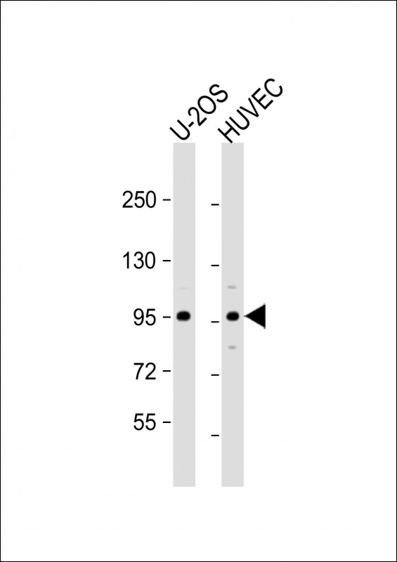
All lanes : Anti-GIT1 Antibody (Center) at 1:2000 dilution Lane 1: U-2OS whole cell lysate Lane 2: HUVEC whole cell lysate Lysates/proteins at 20 µg per lane. Secondary Goat Anti-Rabbit IgG, (H+L), Peroxidase conjugated at 1/10000 dilution. Predicted band size : 84 kDa Blocking/Dilution buffer: 5% NFDM/TBST.

-
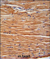
GIT1 Antibody (Center) (Cat. #P34211) immunohistochemistry analysis in formalin fixed and paraffin embedded mouse heart tissue followed by peroxidase conjugation of the secondary antibody and DAB staining. This data demonstrates the use of the GIT1 Antibody (Center) for immunohistochemistry. Clinical relevance has not been evaluated.


 鄂公网安备42018502007531号

