
Mouse Monoclonal Antibody to SMAD1
货号:
P34374
别名:
Mothers against decapentaplegic homolog 1, MAD homolog 1, Mothers against DPP homolog 1, JV4-1, Mad-related protein 1, SMAD family member 1, SMAD 1, Smad1, hSMAD1, Transforming growth factor-beta-signaling protein 1, BSP-1, SMAD1, BSP1, MADH1, MADR1
应用:
WB,IHC,FCM
反应种属:
Human
抗体类型:
Primary antibody
Swissprot:
Q15797
规格:
目录价
在线咨询
Description |
|---|
Transcriptional modulator activated by BMP (bone morphogenetic proteins) type 1 receptor kinase. SMAD1 is a receptor-regulated SMAD (R-SMAD). SMAD1/OAZ1/PSMB4 complex mediates the degradation of the CREBBP/EP300 repressor SNIP1. May act synergistically with SMAD4 and YY1 in bone morphogenetic protein (BMP)-mediated cardiac-specific gene expression. |
Specification |
|
|---|---|
| Aliases | Mothers against decapentaplegic homolog 1, MAD homolog 1, Mothers against DPP homolog 1, JV4-1, Mad-related protein 1, SMAD family member 1, SMAD 1, Smad1, hSMAD1, Transforming growth factor-beta-signaling protein 1, BSP-1, SMAD1, BSP1, MADH1, MADR1 |
| Entrez GeneID | 4086 |
| Swissprot | Q15797 |
| WB Predicted band size | 52.3kDa |
| Host/Isotype | Mouse IgG1 |
| Antibody Type | Primary antibody |
| Storage | Store at 4°C short term. Aliquot and store at -20°C long term. Avoid freeze/thaw cycles. |
| Species Reactivity | Human |
| Immunogen | This SMAD1 antibody is generated from a mouse immunized with a recombinant protein between 20-330 amino acids from human SMAD1. |
Application |
|
|---|---|
| WB | 1/2000 |
| IHC | 1/100-1/500 |
| FCM | 1/25 |
Product Image
-
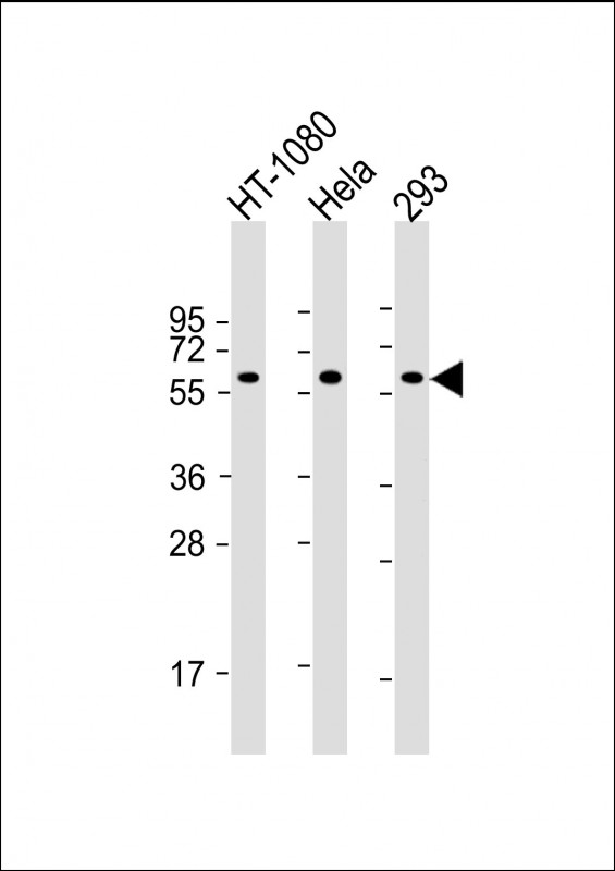
All lanes : Anti-SMAD1 Antibody at 1:2000 dilution Lane 1: HT-1080 whole cell lysate Lane 2: Hela whole cell lysate Lane 3: 293 whole cell lysate Lysates/proteins at 20 µg per lane. Secondary Goat Anti-mouse IgG, (H+L), Peroxidase conjugated at 1/10000 dilution. Predicted band size : 60 kDa Blocking/Dilution buffer: 5% NFDM/TBST.

-
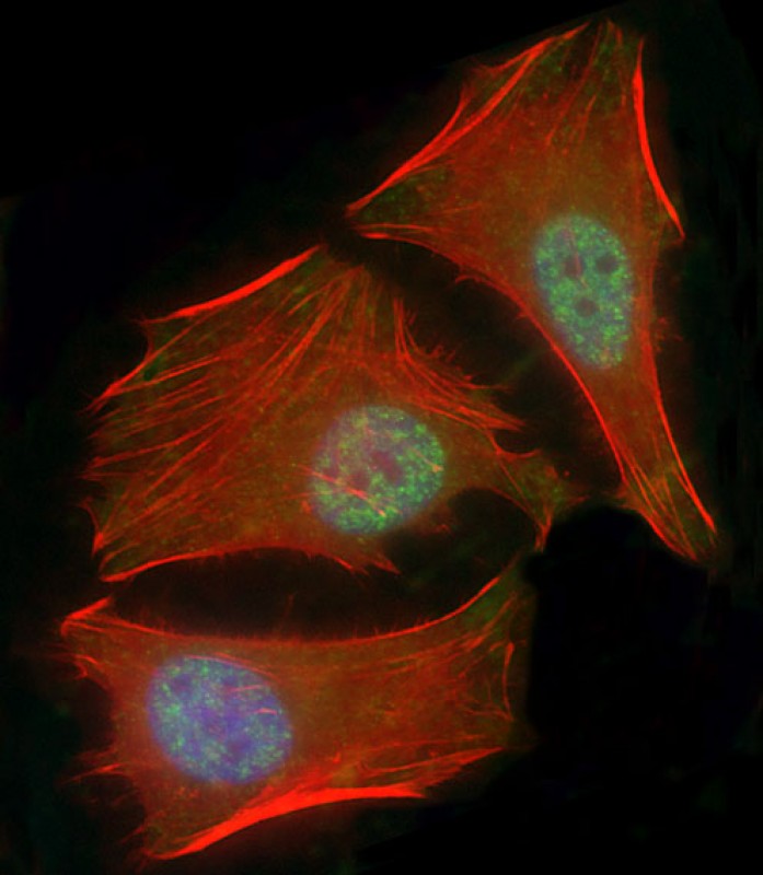
Immunofluorescent analysis of 4% paraformaldehyde-fixed, 0.1% Triton X-100 permeabilized HeLa (human cervical epithelial adenocarcinoma cell line) cells labeling SMAD1 with P34374 at 1/25 dilution, followed by Dylight® 488-conjugated goat anti-mouse IgG secondary antibody at 1/200 dilution (green). Immunofluorescence image showing nucleus and weak cytoplasm staining on HeLa cell line. Cytoplasmic actin is detected with Dylight® 554 Phalloidin at 1/100 dilution (red). The nuclear counter stain is DAPI (blue).

-
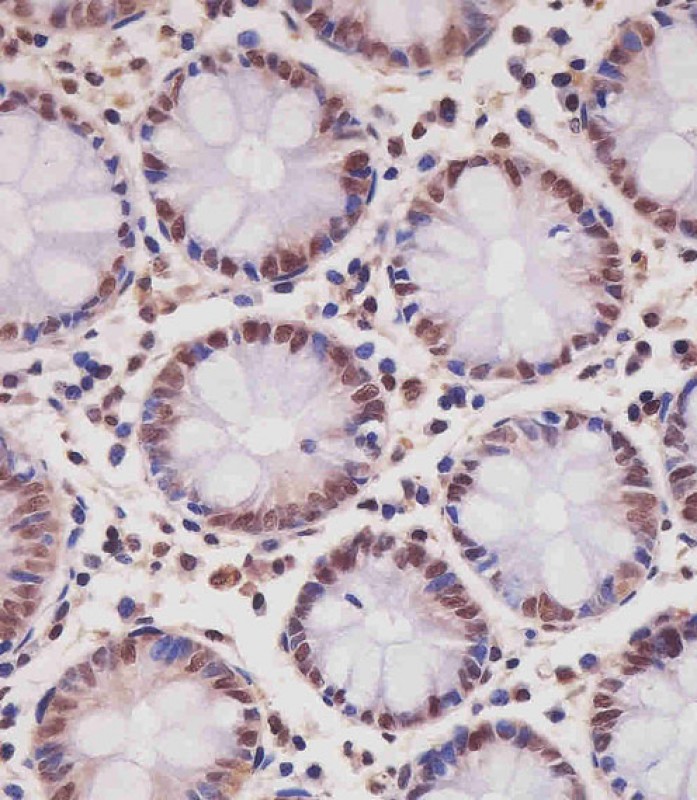
P34374 staining SMAD1 in human colon tissue sections by Immunohistochemistry (IHC-P - paraformaldehyde-fixed, paraffin-embedded sections). Tissue was fixed with formaldehyde and blocked with 3% BSA for 0. 5 hour at room temperature; antigen retrieval was by heat mediation with a citrate buffer (pH6). Samples were incubated with primary antibody (1/25) for 1 hours at 37°C. A undiluted biotinylated goat polyvalent antibody was used as the secondary antibody.

-
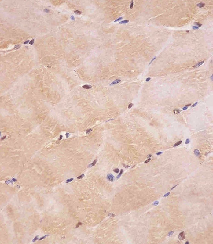
P34374 staining SMAD1 in human skeletal muscle tissue sections by Immunohistochemistry (IHC-P - paraformaldehyde-fixed, paraffin-embedded sections). Tissue was fixed with formaldehyde and blocked with 3% BSA for 0. 5 hour at room temperature; antigen retrieval was by heat mediation with a citrate buffer (pH6). Samples were incubated with primary antibody (1/25) for 1 hours at 37°C. A undiluted biotinylated goat polyvalent antibody was used as the secondary antibody.


 鄂公网安备42018502007531号

