
Mouse Monoclonal Antibody to RAB23
货号:
P34372
别名:
Ras-related protein Rab-23, RAB23
应用:
WB,IF,FCM
反应种属:
Human, Mouse
抗体类型:
Primary antibody
Swissprot:
Q9ULC3
规格:
目录价
在线咨询
Description |
|---|
The protein encoded by this gene belongs to the small GTPase superfamily, Rab family. It may be involved in small GTPase mediated signal transduction and intracellular protein transportation. Alternative splicing occurs at this locus and two transcript variants encoding the same protein have been identified. |
Specification |
|
|---|---|
| Aliases | Ras-related protein Rab-23, RAB23 |
| Entrez GeneID | 51715 |
| Swissprot | Q9ULC3 |
| WB Predicted band size | 26.7kDa |
| Host/Isotype | Mouse IgG1 |
| Antibody Type | Primary antibody |
| Storage | Store at 4°C short term. Aliquot and store at -20°C long term. Avoid freeze/thaw cycles. |
| Species Reactivity | Human, Mouse |
| Immunogen | Purified His-tagged RAB23 protein(Fragment) was used to produced this monoclonal antibody. |
| Formulation | Purified antibody in TBS with 0.05% sodium azide. |
Application |
|
|---|---|
| WB | 1/2000 |
| IF | 1/25 |
| FCM | 1/25 |
Product Image
-
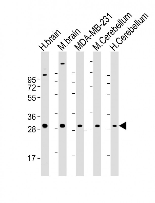
All lanes : Anti-RAB23 Antibody at 1:2000 dilution Lane 1: human brain lysate Lane 2: mouse brain lysate Lane 3: MDA-MB-231 whole cell lysate Lane 4: mouse Cerebellum lysate Lane 5: human Cerebellum lysate Lysates/proteins at 20 µg per lane. Secondary Goat Anti-mouse IgG, (H+L), Peroxidase conjugated at 1/10000 dilution. Predicted band size : 27 kDa Blocking/Dilution buffer: 5% NFDM/TBST.

-
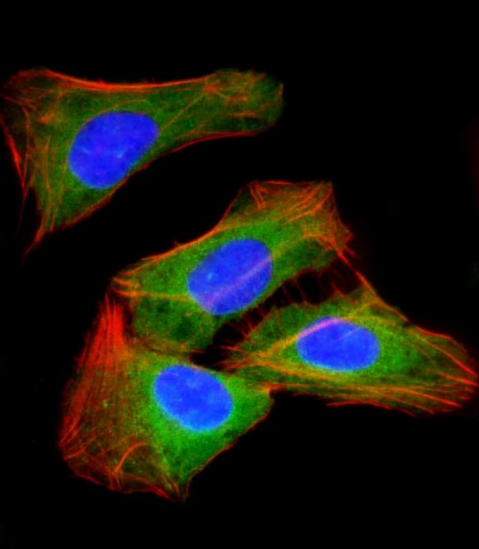
Immunofluorescent analysis of 4% paraformaldehyde-fixed, 0.1% Triton X-100 permeabilized U-2 OS (human osteosarcoma cell line) cells labeling RAB23 with AM2026a at 1/25 dilution, followed by Dylight® 488-conjugated goat anti-mouse IgG secondary antibody at 1/200 dilution (green). Immunofluorescence image showing cytoplasm staining on U-2 OS cell line. Cytoplasmic actin is detected with Dylight® 554 Phalloidin at 1/100 dilution (red).The nuclear counter stain is DAPI (blue).

-
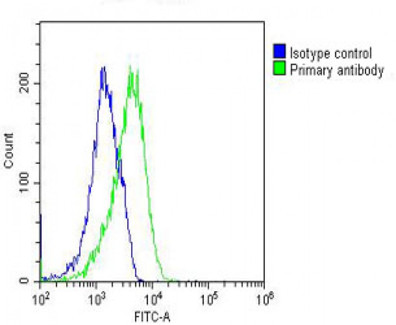
Overlay histogram showing U-2 OS cells stained with AM2026a(green line). The cells were fixed with 2% paraformaldehyde (10 min) and then permeabilized with 90% methanol for 10 min. The cells were then icubated in 2% bovine serum albumin to block non-specific protein-protein interactions followed by the antibody (AM2026a, 1:25 dilution) for 60 min at 37ºC. The secondary antibody used was Goat-Anti-Mouse IgG, DyLight® 488 Conjugated Highly Cross-Adsorbed(NH174309) at 1/200 dilution for 40 min at 37ºC. Isotype control antibody (blue line) was mouse IgG1(1μg/1x10^6 cells) used under the same conditions. Acquisition of >10, 000 events was performed.


 鄂公网安备42018502007531号

