
Mouse Monoclonal Antibody to HIF1AN
货号:
P34411
别名:
Hypoxia-inducible factor 1-alpha inhibitor, 1.14.11.30, 1.14.11.n4, Factor inhibiting HIF-1, FIH-1, Hypoxia-inducible factor asparagine hydroxylase, HIF1AN, FIH1
应用:
WB,IF
反应种属:
Human
抗体类型:
Primary antibody
Swissprot:
Q9NWT6
规格:
目录价
在线咨询
Description |
|---|
Hydroxylates HIF-1 alpha at 'Asp-803' in the C-terminal transactivation domain (CAD). Functions as an oxygen sensor and, under normoxic conditions, the hydroxylation prevents interaction of HIF-1 with transcriptional coactivators including Cbp/p300- interacting transactivator. Involved in transcriptional repression through interaction with HIF1A, VHL and histone deacetylases. Hydroxylates specific Asn residues within ankyrin repeat domains (ARD) of NFKB1, NFKBIA, NOTCH1, ASB4, PPP1R12A and several other ARD-containing proteins. Also hydroxylates Asp and His residues within ARDs of ANK1 and TNKS2, respectively. Negatively regulates NOTCH1 activity, accelerating myogenic differentiation. Positively regulates ASB4 activity, promoting vascular differentiation. |
Specification |
|
|---|---|
| Aliases | Hypoxia-inducible factor 1-alpha inhibitor, 1.14.11.30, 1.14.11.n4, Factor inhibiting HIF-1, FIH-1, Hypoxia-inducible factor asparagine hydroxylase, HIF1AN, FIH1 |
| Entrez GeneID | 55662 |
| Swissprot | Q9NWT6 |
| WB Predicted band size | 40.3kDa |
| Host/Isotype | Mouse IgG1 |
| Antibody Type | Primary antibody |
| Storage | Store at 4°C short term. Aliquot and store at -20°C long term. Avoid freeze/thaw cycles. |
| Species Reactivity | Human |
| Immunogen | This HIF1AN antibody is generated from a mouse immunized with a KLH conjugated synthetic peptide between 1-349 amino acids from human HIF1AN. |
Application |
|
|---|---|
| WB | 1/2000 |
| IF | 1/25 |
Product Image
-
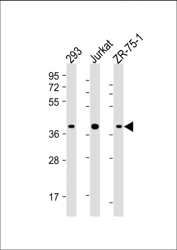
All lanes : Anti-HIF1AN Antibody (C-term) at 1:4000 dilution Lane 1: 293 whole cell lysate Lane 2: Jurkat whole cell lysate Lane 3: ZR-75-1 whole cell lysate Lysates/proteins at 20 µg per lane. Secondary Goat Anti-mouse IgG, (H+L), Peroxidase conjugated at 1/10000 dilution. Predicted band size : 40 kDa Blocking/Dilution buffer: 5% NFDM/TBST.

-
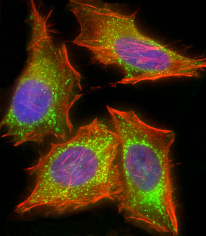
Immunofluorescent analysis of 4% paraformaldehyde-fixed, 0.1% Triton X-100 permeabilized HeLa (human cervical epithelial adenocarcinoma cell line) cells labeling HIF1AN with P34411 at 1/25 dilution, followed by Dylight® 488-conjugated goat anti-mouse IgG secondary antibody at 1/200 dilution (green). Immunofluorescence image showing mitochondrion staining on HeLa cell line. Cytoplasmic actin is detected with Dylight® 554 Phalloidin at 1/100 dilution (red). The nuclear counter stain is DAPI (blue).


 鄂公网安备42018502007531号

