

Rabbit Polyclonal Antibody to PLEC
货号:
P34436
别名:
Plectin, PCN, PLTN, Hemidesmosomal protein 1, HD1, Plectin-1, PLEC, PLEC1
应用:
WB,IF
反应种属:
Human, Mouse, Rat
抗体类型:
Primary antibody
Swissprot:
Q15149
规格:
目录价
在线咨询
Description |
|---|
Interlinks intermediate filaments with microtubules and microfilaments and anchors intermediate filaments to desmosomes or hemidesmosomes. Could also bind muscle proteins such as actin to membrane complexes in muscle. May be involved not only in the filaments network, but also in the regulation of their dynamics. Structural component of muscle. Isoform 9 plays a major role in the maintenance of myofibers integrity. |
Specification |
|
|---|---|
| Aliases | Plectin, PCN, PLTN, Hemidesmosomal protein 1, HD1, Plectin-1, PLEC, PLEC1 |
| Entrez GeneID | 5339 |
| Swissprot | Q15149 |
| WB Predicted band size | 531.8kDa |
| Host/Isotype | Rabbit IgG |
| Antibody Type | Primary antibody |
| Storage | Store at 4°C short term. Aliquot and store at -20°C long term. Avoid freeze/thaw cycles. |
| Species Reactivity | Human, Mouse, Rat |
| Immunogen | This PLEC antibody is generated from a rabbit immunized with a KLH conjugated synthetic peptide between 4241-4275 amino acids from human PLEC. |
Application |
|
|---|---|
| WB | 1/1000 |
| IF | 1/25 |
Product Image
-
Anti-PLEC Antibody (C-Term) at 1:1000 dilution + U-2OS whole cell lysate Lysates/proteins at 20 µg per lane. Secondary Goat Anti-Rabbit IgG, (H+L), Peroxidase conjugated at 1/10000 dilution. Predicted band size : 532 kDa Blocking/Dilution buffer: 5% NFDM/TBST.

-
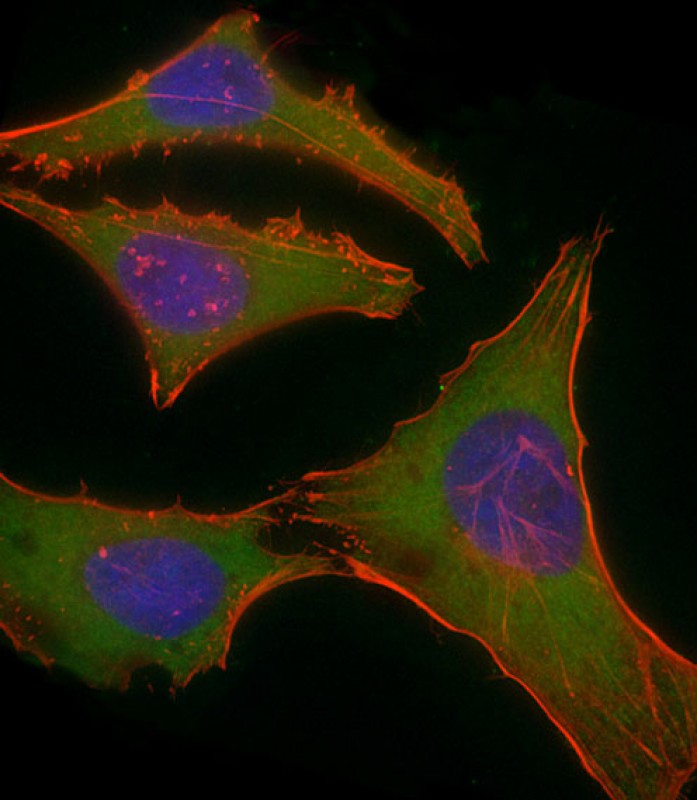
Immunofluorescent analysis of 4% paraformaldehyde-fixed, 0.1% Triton X-100 permeabilized HeLa (human cervical epithelial adenocarcinoma cell line) cells labeling PLEC with P34436 at 1/25 dilution, followed by Dylight® 488-conjugated goat anti-rabbit IgG secondary antibody at 1/200 dilution (green). Immunofluorescence image showing cytoplasm staining on HeLa cell line. Cytoplasmic actin is detected with Dylight® 554 Phalloidin at 1/100 dilution (red). The nuclear counter stain is DAPI (blue).


 鄂公网安备42018502007531号

