
Rabbit Polyclonal Antibody to NOX1
货号:
P34662
别名:
NADPH oxidase 1, NOX-1, 1---, Mitogenic oxidase 1, MOX-1, NADH/NADPH mitogenic oxidase subunit P65-MOX, NOH-1, NOX1, MOX1, NOH1
应用:
WB,IHC
反应种属:
Human
抗体类型:
Primary antibody
Swissprot:
Q9Y5S8
规格:
目录价
在线咨询
Description |
|---|
Voltage-gated proton (hydrogen) channels play an important role in cellular defense against acidic stress. They are unique among ion channels with respect to their extremely high selectivity, marked temperature dependence, and unitary conductance, which is 3 orders of magnitude lower than that of most other ion channels. NOX1 is a homolog of the catalytic subunit of the superoxide-generating NADPH oxidase of phagocytes, gp91phox. Two transcript variants encoding different isoforms have been found for this gene. |
Specification |
|
|---|---|
| Aliases | NADPH oxidase 1, NOX-1, 1---, Mitogenic oxidase 1, MOX-1, NADH/NADPH mitogenic oxidase subunit P65-MOX, NOH-1, NOX1, MOX1, NOH1 |
| Entrez GeneID | 27035 |
| Swissprot | Q9Y5S8 |
| WB Predicted band size | 64.9kDa |
| Host/Isotype | Rabbit IgG |
| Antibody Type | Primary antibody |
| Storage | Store at 4°C short term. Aliquot and store at -20°C long term. Avoid freeze/thaw cycles. |
| Species Reactivity | Human |
| Immunogen | This NOX1 antibody is generated from rabbits immunized with a KLH conjugated synthetic peptide between 243-271 amino acids from the Central region of human NOX1. |
| Formulation | Purified antibody in PBS with 0.05% sodium azide. |
Application |
|
|---|---|
| WB | 1/1000 |
| IHC | 1/500 |
Product Image
-
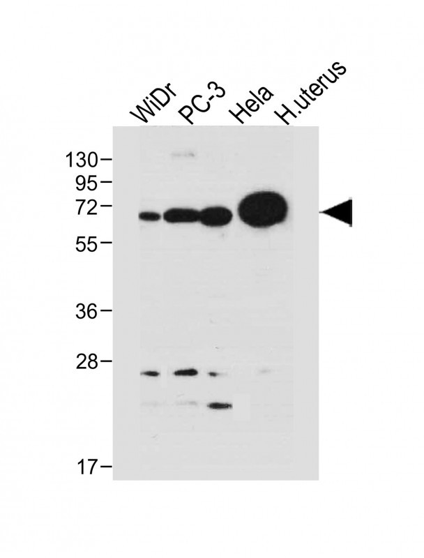
All lanes : Anti-NOX1 Antibody (Center) at 1:1000 dilution Lane 1: WiDr whole cell lysate Lane 2: PC-3 whole cell lysate Lane 3: Hela whole cell lysate Lane 4: human uterus tissue lysate Lysates/proteins at 20 µg per lane. Secondary Goat Anti-Rabbit IgG, (H+L), Peroxidase conjugated at 1/10000 dilution. Predicted band size : 65 kDa Blocking/Dilution buffer: 5% NFDM/TBST.

-
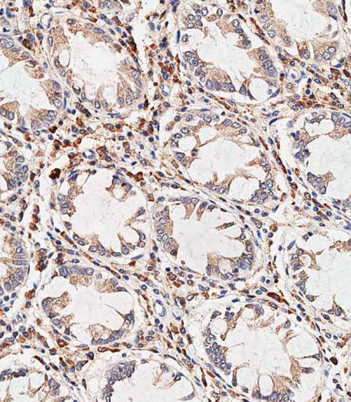
Immunohistochemical analysis of paraffin-embedded human colon tissue using P34662 performed on the Leica® BOND RXm. Tissue was fixed with formaldehyde at room temperature, antigen retrieval was by heat mediation with a EDTA buffer (pH9. 0). Samples were incubated with primary antibody(1:500) for 1 hours at room temperature. A undiluted biotinylated CRF Anti-Polyvalent HRP Polymer antibody was used as the secondary antibody.

-
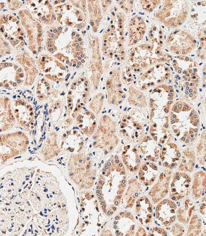
Immunohistochemical analysis of paraffin-embedded human kidney tissue using P34662 performed on the Leica® BOND RXm. Tissue was fixed with formaldehyde at room temperature, antigen retrieval was by heat mediation with a EDTA buffer (pH9. 0). Samples were incubated with primary antibody(1:500) for 1 hours at room temperature. A undiluted biotinylated CRF Anti-Polyvalent HRP Polymer antibody was used as the secondary antibody.


 鄂公网安备42018502007531号

