
Rabbit Polyclonal Antibody to PHO1 (N-term)
货号:
P34661
别名:
DNA dC->dU-editing enzyme APOBEC-3A, A3A, 354-, Phorbolin-1, APOBEC3A
应用:
WB,IHC
反应种属:
Human
抗体类型:
Primary antibody
Swissprot:
P31941
规格:
目录价
在线咨询
Description |
|---|
PHO1 a member of the cytidine deaminase gene family. The PHO1 gene is one of seven related genes or pseudogenes found in a cluster, thought to result from gene duplication, on chromosome 22. Members of the cluster encode proteins that are structurally and functionally related to the C to U RNA-editing cytidine deaminase APOBEC1. It is thought that the proteins may be RNA editing enzymes and have roles in growth or cell cycle control. This gene encodes a protein that lacks the zinc binding activity and may be an expressed pseudogene. |
Specification |
|
|---|---|
| Aliases | DNA dC->dU-editing enzyme APOBEC-3A, A3A, 354-, Phorbolin-1, APOBEC3A |
| Entrez GeneID | 100913187;200315 |
| Swissprot | P31941 |
| WB Predicted band size | 23.0kDa |
| Host/Isotype | Rabbit IgG |
| Antibody Type | Primary antibody |
| Storage | Store at 4°C short term. Aliquot and store at -20°C long term. Avoid freeze/thaw cycles. |
| Species Reactivity | Human |
| Immunogen | This PHO1 antibody is generated from rabbits immunized with a KLH conjugated synthetic peptide between 1-30 amino acids from the N-terminal region of human PHO1. |
| Formulation | Purified antibody in PBS with 0.05% sodium azide,1%BSA and 50% glycerol.prepared by Saturated Ammonium Sulfate (SAS) . |
Application |
|
|---|---|
| WB | 1/2000 |
| IHC | 1/100-1/500 |
Product Image
-
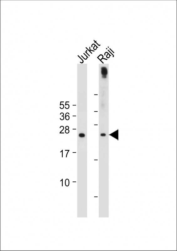
All lanes : Anti-hPHO1-M1 at 1:2000 dilution Lane 1: Jurkat whole cell lysate Lane 2: Raji whole cell lysate Lysates/proteins at 20 µg per lane. Secondary Goat Anti-Rabbit IgG, (H+L), Peroxidase conjugated at 1/10000 dilution. Predicted band size : 23 kDa Blocking/Dilution buffer: 5% NFDM/TBST.

-
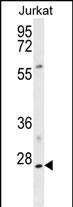
Western blot analysis of hPHO1-M1 (Cat. #P34661) in Jurkat cell line lysates (35ug/lane). PHO1 (arrow) was detected using the purified Pab.

-
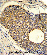
Formalin-fixed and paraffin-embedded human prostate carcinoma reacted with PHO1 Antibody (N-term), which was peroxidase-conjugated to the secondary antibody, followed by DAB staining. This data demonstrates the use of this antibody for immunohistochemistry; clinical relevance has not been evaluated.


 鄂公网安备42018502007531号

