
Mouse Monoclonal Antibody to SET07
货号:
P34848
别名:
N-lysine methyltransferase SETD8, 211-, H4-K20-HMTase SETD8, Histone-lysine N-methyltransferase SETD8, Lysine N-methyltransferase 5A, PR/SET domain-containing protein 07, PR-Set7, PR/SET07, SET domain-containing protein 8, SETD8, KMT5A, PRSET7, SET07, SET
应用:
WB,IHC,IF
反应种属:
Human
抗体类型:
Primary antibody
Swissprot:
Q9NQR1
规格:
目录价
在线咨询
Specification |
|
|---|---|
| Aliases | N-lysine methyltransferase SETD8, 211-, H4-K20-HMTase SETD8, Histone-lysine N-methyltransferase SETD8, Lysine N-methyltransferase 5A, PR/SET domain-containing protein 07, PR-Set7, PR/SET07, SET domain-containing protein 8, SETD8, KMT5A, PRSET7, SET07, SET8 |
| Entrez GeneID | 387893 |
| Swissprot | Q9NQR1 |
| WB Predicted band size | 42.9kDa |
| Host/Isotype | Mouse IgG1 |
| Antibody Type | Primary antibody |
| Storage | Store at 4°C short term. Aliquot and store at -20°C long term. Avoid freeze/thaw cycles. |
| Species Reactivity | Human |
| Immunogen | This SET07 antibody was raised using purified recombinant GST fusion protein encoding the N-terminal region of human SET07. |
| Formulation | Purified antibody in PBS with 0.05% sodium azide. |
Application |
|
|---|---|
| WB | 1/2000 |
| IHC | 1/100-1/500 |
| IF | 1/10-1/50 |
Product Image
-
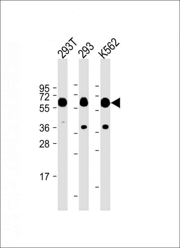
All lanes :SET07 Antibody at 1:2000 dilution Lane 1: 293T whole cell lysate Lane 2: 293 whole cell lysate Lane 3: K562 whole cell lysate Lysates/proteins at 20 µg per lane. Secondary Goat Anti-Rabbit IgG, (H+L), Peroxidase conjugated at 1/10000 dilution. Predicted band size : 43 kDa Blocking/Dilution buffer: 5% NFDM/TBST.

-
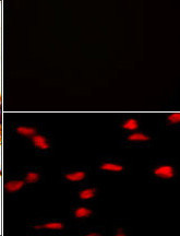
(TOP)Negative control of hela cells without PE-conjugated goat anti-mouse lgG (whole molecule).PE-conjugated goat anti-mouse lgG emits red fluorescence. ( Bottom)Immunofluorescence analysis of SET07 Antibody in HeLa cells. 0.025 mg/ml primary antibody was followed by PE-conjugated goat anti-mouse lgG (whole molecule).

-
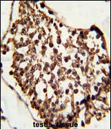
Formalin-fixed and paraffin-embedded human testis tissue reacted with SET07 Antibody, which was peroxidase-conjugated to the secondary antibody, followed by DAB staining. This data demonstrates the use of this antibody for immunohistochemistry; clinical relevance has not been evaluated.


 鄂公网安备42018502007531号

