
Rabbit Polyclonal Antibody to ND5
货号:
P34943
别名:
NADH-ubiquinone oxidoreductase chain 5, NADH dehydrogenase subunit 5, MT-ND5, MTND5, NADH5, ND5
应用:
WB,IHC,FCM
反应种属:
Human
抗体类型:
Primary antibody
Swissprot:
P03915
规格:
目录价
在线咨询
Description |
|---|
Core subunit of the mitochondrial membrane respiratory chain NADH dehydrogenase (Complex I) that is believed to belong to the minimal assembly required for catalysis. Complex I functions in the transfer of electrons from NADH to the respiratory chain. The immediate electron acceptor for the enzyme is believed to be ubiquinone. |
Specification |
|
|---|---|
| Aliases | NADH-ubiquinone oxidoreductase chain 5, NADH dehydrogenase subunit 5, MT-ND5, MTND5, NADH5, ND5 |
| Entrez GeneID | 4540 |
| Swissprot | P03915 |
| WB Predicted band size | 67.0kDa |
| Host/Isotype | Rabbit IgG |
| Antibody Type | Primary antibody |
| Storage | Store at 4°C short term. Aliquot and store at -20°C long term. Avoid freeze/thaw cycles. |
| Species Reactivity | Human |
| Immunogen | This ND5 antibody is generated from rabbits immunized with a KLH conjugated synthetic peptide between 544-570 amino acids from the C-terminal region of human ND5. |
| Formulation | Purified antibody in PBS with 0.05% sodium azide. |
Application |
|
|---|---|
| WB | 1/1000 |
| IHC | 1/100-1/500 |
| FCM | 1/10-1/50 |
Product Image
-
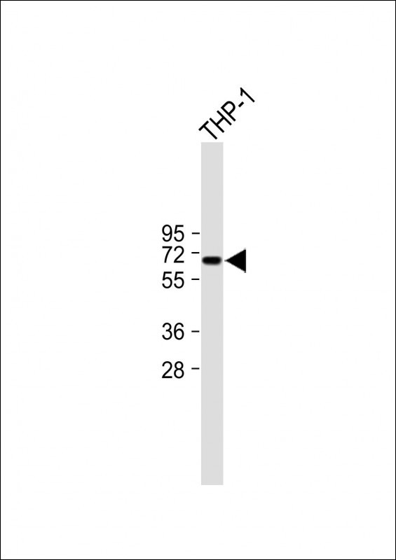
All lanes : Anti-ND5 Antibody (C-term) at 1:1000 dilution Lane 1: THP-1 whole cell lysate Lysates/proteins at 20 µg per lane. Secondary Goat Anti-Rabbit IgG, (H+L), Peroxidase conjugated (ASP1615) at 1/15000 dilution. Observed band size : 67kDa Blocking/Dilution buffer: 5% NFDM/TBST.

-
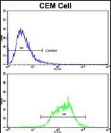
Flow cytometric analysis of CEM cells using ND5 Antibody (C-term)(bottom histogram) compared to a negative control cell (top histogram). FITC-conjugated goat-anti-rabbit secondary antibodies were used for the analysis.

-
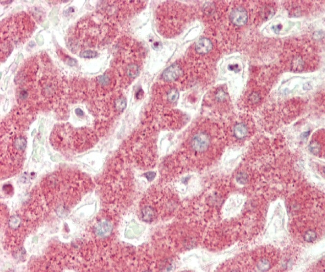
Formalin-fixed and paraffin-embedded H.liver tissue reacted with ND5 Antibody (C-term) (Cat#P34943).

-
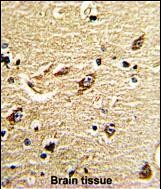
Formalin-fixed and paraffin-embedded human brain tissue reacted with ND5 Antibody (C-term), which was peroxidase-conjugated to the secondary antibody, followed by DAB staining. This data demonstrates the use of this antibody for immunohistochemistry; clinical relevance has not been evaluated.


 鄂公网安备42018502007531号

