
Rabbit Polyclonal Antibody to MVD
货号:
P34941
别名:
Diphosphomevalonate decarboxylase, Mevalonate (diphospho)decarboxylase, MDDase, Mevalonate pyrophosphate decarboxylase, MVD, MPD
应用:
WB,IHC,FCM
反应种属:
Human, Mouse, Rat
抗体类型:
Primary antibody
Swissprot:
P53602
规格:
目录价
在线咨询
Description |
|---|
The enzyme mevalonate pyrophosphate decarboxylase catalyzes the conversion of mevalonate pyrophosphate into isopentenyl pyrophosphate in one of the early steps in cholesterol biosynthesis. It decarboxylates and dehydrates its substrate while hydrolyzing ATP. |
Specification |
|
|---|---|
| Aliases | Diphosphomevalonate decarboxylase, Mevalonate (diphospho)decarboxylase, MDDase, Mevalonate pyrophosphate decarboxylase, MVD, MPD |
| Entrez GeneID | 4597 |
| Swissprot | P53602 |
| WB Predicted band size | 43.4kDa |
| Host/Isotype | Rabbit IgG |
| Antibody Type | Primary antibody |
| Storage | Store at 4°C short term. Aliquot and store at -20°C long term. Avoid freeze/thaw cycles. |
| Species Reactivity | Human, Mouse, Rat |
| Immunogen | This MVD antibody is generated from rabbits immunized with a KLH conjugated synthetic peptide between 164-193 amino acids from the Central region of human MVD. |
| Formulation | Purified antibody in PBS with 0.05% sodium azide,1%BSA and 50% glycerol.prepared by Saturated Ammonium Sulfate (SAS) . |
Application |
|
|---|---|
| WB | 1/1000 |
| IHC | 1/100-1/500 |
| FCM | 1/10-1/50 |
Product Image
-
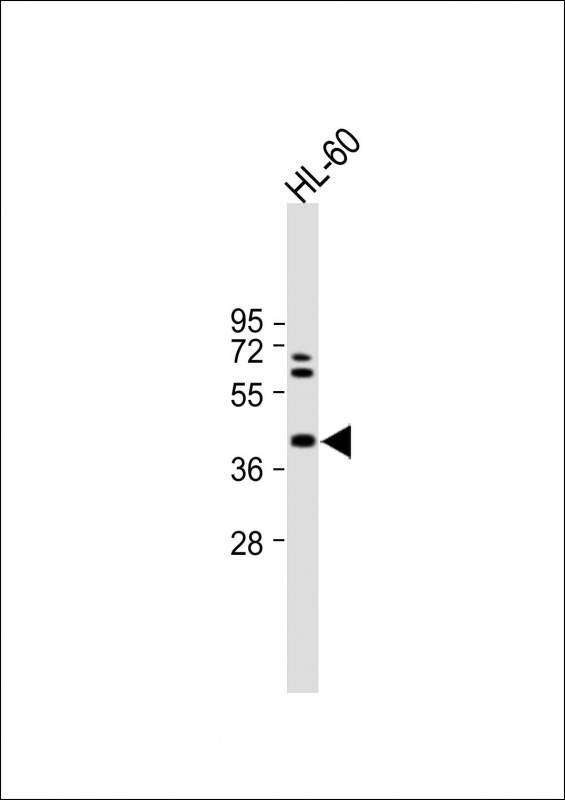
All lanes : Anti-MVD Antibody (Center) at 1:1000 dilution Lane 1: HL-60 whole cell lysate Lysates/proteins at 20 µg per lane. Secondary Goat Anti-Rabbit IgG, (H+L), Peroxidase conjugated (ASP1615) at 1/15000 dilution. Observed band size : 43kDa Blocking/Dilution buffer: 5% NFDM/TBST.

-
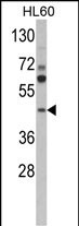
Western blot analysis of MVD Antibody (Center)(Cat. #P34941) in HL60 cell line lysates (35ug/lane). MVD (arrow) was detected using the purified Pab.

-
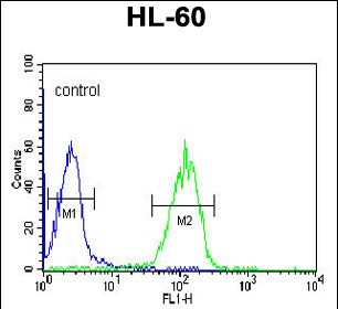
MVD Antibody (Center) (Cat. #P34941) flow cytometric analysis of HL-60 cells (right histogram) compared to a negative control cell (left histogram).FITC-conjugated goat-anti-rabbit secondary antibodies were used for the analysis.

-
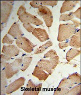
Formalin-fixed and paraffin-embedded human Skeletal muscle reacted with MVD Antibody (Center), which was peroxidase-conjugated to the secondary antibody, followed by DAB staining. This data demonstrates the use of this antibody for immunohistochemistry; clinical relevance has not been evaluated.


 鄂公网安备42018502007531号

