
Mouse Monoclonal Antibody to ALPG
货号:
32269
别名:
GCAP; ALPPL; ALPPL2
应用:
WB,IHC,IF,FCM
反应种属:
Human
抗体类型:
Primary antibody
Swissprot:
P10696
规格:
目录价
在线咨询
Description |
|---|
There are at least four distinct but related alkaline phosphatases: intestinal, placental, placental-like, and liver/bone/kidney (tissue non-specific). The product of this gene is a membrane bound glycosylated enzyme, localized to testis, thymus and certain germ cell tumors, that is closely related to both the placental and intestinal forms of alkaline phosphatase. |
References |
|---|
| 1.Cancer Res. 2020 Oct 15;80(20):4552-4564. 2.Cell Rep. 2020 Mar 17;30(11):3917-3931.e5. |
Specification |
|
|---|---|
| Aliases | GCAP; ALPPL; ALPPL2 |
| Entrez GeneID | 251 |
| Swissprot | P10696 |
| clone | 3A2B7 |
| WB Predicted band size | 57.3kDa |
| Host/Isotype | Mouse IgG2b |
| Antibody Type | Primary antibody |
| Storage | Store at 4°C short term. Aliquot and store at -20°C long term. Avoid freeze/thaw cycles. |
| Species Reactivity | Human |
| Immunogen | Purified recombinant fragment of human ALPG (AA: 170-285) expressed in E. Coli. |
| Formulation | Purified antibody in PBS with 0.05% sodium azide |
Application |
|
|---|---|
| WB | 1/500 - 1/2000 |
| IHC | 1/200 - 1/1000 |
| IF | 1/50 - 1/200 |
| FCM | 1/200 - 1/400 |
| ELISA | 1/10000 |
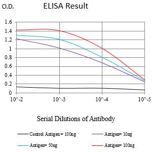
Product Image
-
Black line: Control Antigen (100 ng);Purple line: Antigen (10ng); Blue line: Antigen (50 ng); Red line:Antigen (100 ng)

-
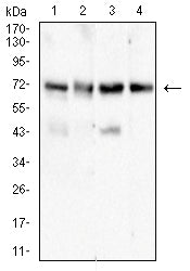
Western blot analysis using ALPG mouse mAb against SK-OV-3 (1), Hela (2),HepG2 (3), and A431 (4) cell lysate.

-
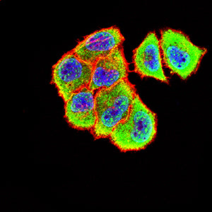
Immunofluorescence analysis of Hela cells using ALPG mouse mAb (green). Blue: DRAQ5 fluorescent DNA dye. Red: Actin filaments have been labeled with Alexa Fluor- 555 phalloidin. Secondary antibody from Fisher (Cat#: 35503)

-
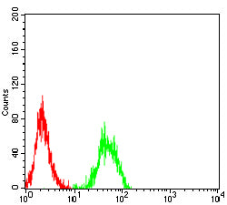
Flow cytometric analysis of Jurkat cells using ALPG mouse mAb (green) and negative control (red).

-
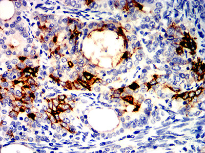
Immunohistochemical analysis of paraffin-embedded human cervical cancer tissues using ALPG mouse mAb with DAB staining.

-
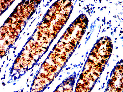
Immunohistochemical analysis of paraffin-embedded human rectum cancer tissues using ALPG mouse mAb with DAB staining.


 鄂公网安备42018502007531号

