

Mouse Monoclonal Antibody to C-CBL
货号:
30250
别名:
CBL; CBL2; NSLL; C-CBL; RNF55
应用:
WB,IHC,IF,FCM
反应种属:
Human,Mouse,Rat,Monkey,Rabbit
抗体类型:
Primary antibody
Swissprot:
P22681
规格:
目录价
在线咨询
Description |
|---|
The cbl oncogene was first identified as part of a transforming retrovirus which induces mouse pre-B and pro-B cell lymphomas. As an adaptor protein for receptor protein-tyrosine kinases, it positively regulates receptor protein-tyrosine kinase ubiquitination in a manner dependent upon its variant SH2 and RING finger domains. Ubiquitination of receptor protein-tyrosine kinases terminates signaling by marking active receptors for degradation. |
References |
|---|
| 1. Blood. 2009 Aug 27;114(9):1859-63. 2. Cell Res. 2009 Aug;19(8):950-61. 3. Nature. 2009 Aug 13;460(7257):904-8. |
Specification |
|
|---|---|
| Aliases | CBL; CBL2; NSLL; C-CBL; RNF55 |
| Entrez GeneID | 867 |
| Swissprot | P22681 |
| clone | 3B12 |
| WB Predicted band size | 120kDa |
| Host/Isotype | Mouse IgG1 |
| Antibody Type | Primary antibody |
| Storage | Store at 4°C short term. Aliquot and store at -20°C long term. Avoid freeze/thaw cycles. |
| Species Reactivity | Human,Mouse,Rat,Monkey,Rabbit |
| Immunogen | Purified recombinant fragment of human C-CBL expressed in E. Coli. |
| Formulation | Purified antibody in PBS with 0.05% sodium azide. |
Application |
|
|---|---|
| WB | 1/500 - 1/2000 |
| IHC | 1/100 - 1/500 |
| IF | 1/50 - 1/500 |
| FCM | 1/200 - 1/400 |
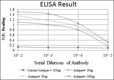
| ELISA | 1/10000 |
Product Image
-
Red: Control Antigen (100ng); Purple: Antigen (10ng); Green: Antigen (50ng); Blue: Antigen (100ng);

-
Western blot analysis using C-CBL mouse mAb against RAJI (1), RAW264.7 (2), K562 (3), SKBR-3 (4), 3T3-L1 (5), THP-1 (6) and PC-12 (7) cell lysate.

-
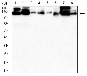
Western blot analysis using C-CBL mouse mAb against PC-12(1)Raw264.7(2)NIH/3T3(3)NRK(4)C2C12(5)C6(6)F9(7)COS-7(8) cell lysate.

-
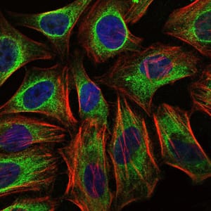
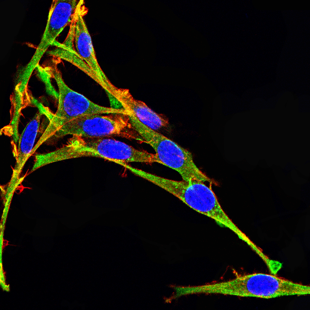
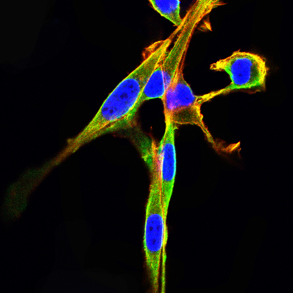
Immunofluorescence analysis of Hela cells using C-CBL mouse mAb (green). Blue: DRAQ5 fluorescent DNA dye. Red: Actin filaments have been labeled with Alexa Fluor-555 phalloidin.

-
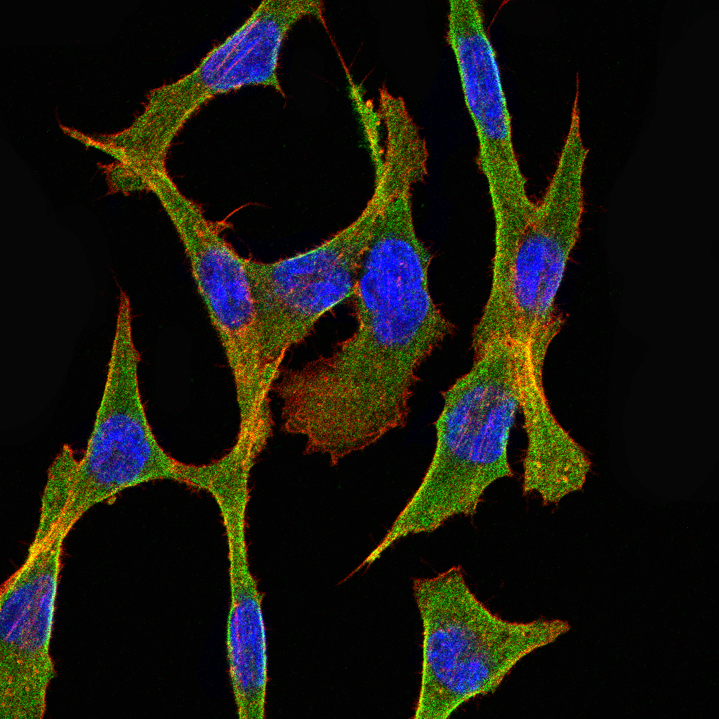
Immunofluorescence analysis of COS7 cells using C-CBL mouse mAb (green). Blue: DRAQ5 fluorescent DNA dye. Red: Actin filaments have been labeled with Alexa Fluor- 555 phalloidin.

-
Immunofluorescence analysis of PC-12 cells using C-CBL mouse mAb (green). Blue: DRAQ5 fluorescent DNA dye. Red: Actin filaments have been labeled with Alexa Fluor- 555 phalloidin.

-
Immunofluorescence analysis of NIH/3T3 cells using C-CBL mouse mAb (green). Blue: DRAQ5 fluorescent DNA dye. Red: Actin filaments have been labeled with Alexa Fluor- 555 phalloidin.

-
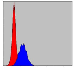
Flow cytometric analysis of MCF-7 cells using C-CBL mouse mAb (blue) and negative control (red).

-
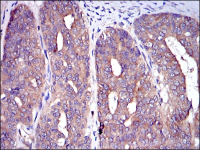
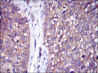
Immunohistochemical analysis of paraffin-embedded human ovarian cancer tissues using C-CBL mouse mAb with DAB staining.

-
Immunohistochemical analysis of paraffin-embedded human bladder cancer tissues using C-CBL mouse mAb with DAB staining.

-
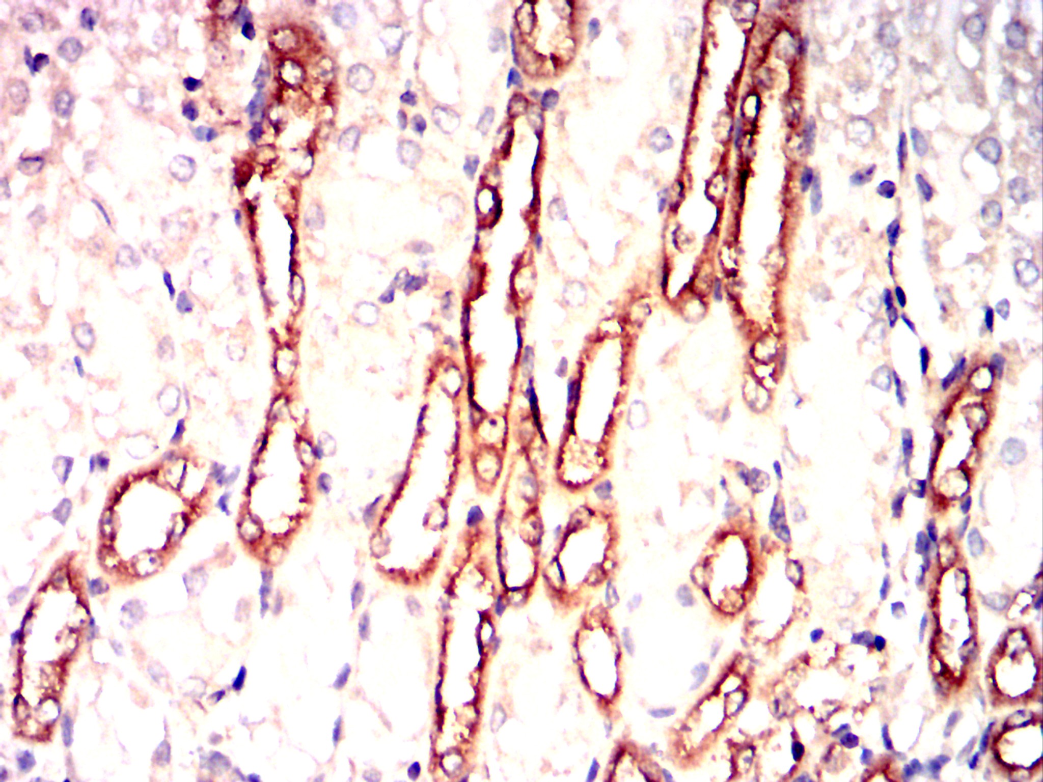
Immunohistochemical analysis of paraffin-embedded Mouse kidney using C-CBL mouse mAb with DAB staining.

-
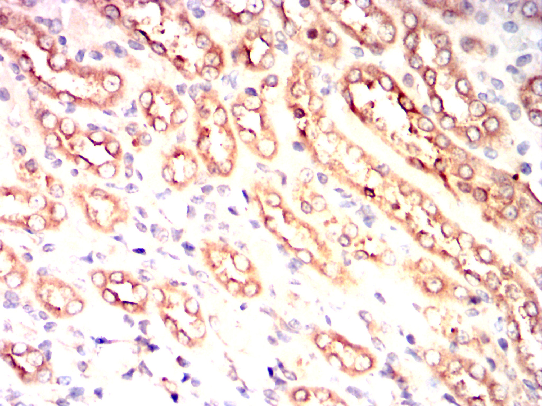
Immunohistochemical analysis of paraffin-embedded Rat kidney using C-CBL mouse mAb with DAB staining.

-
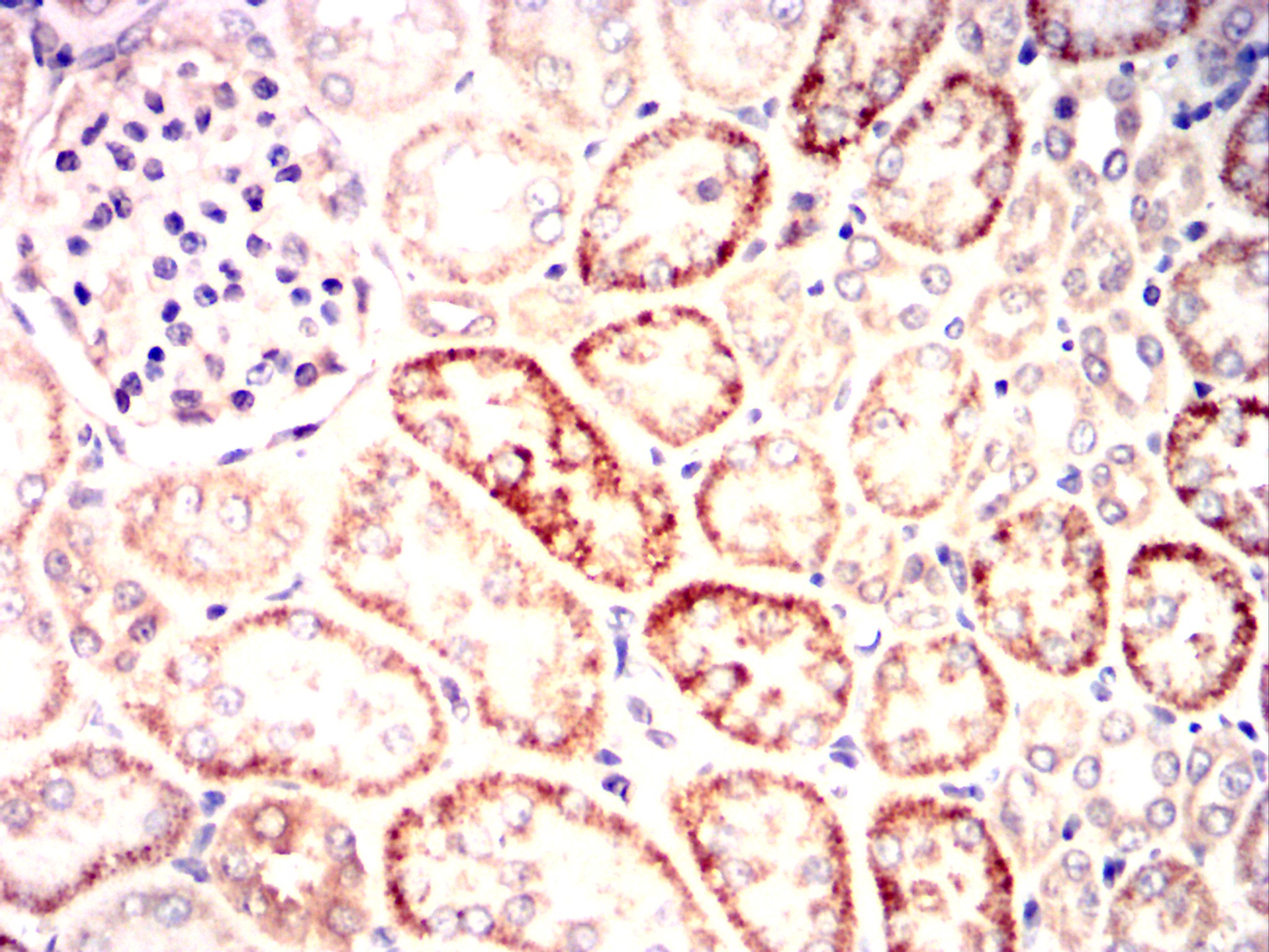
Immunohistochemical analysis of paraffin-embedded Rabbit kidney using C-CBL mouse mAb with DAB staining.


 鄂公网安备42018502007531号

